

Chondroblastoma Located in the Anterior Skull Base: A Case Report and Comprehensive Literature Review
- PMID: 40822611
- PMCID: PMC12352982
- DOI: 10.1155/crot/9368865
Chondroblastoma Located in the Anterior Skull Base: A Case Report and Comprehensive Literature Review
Abstract
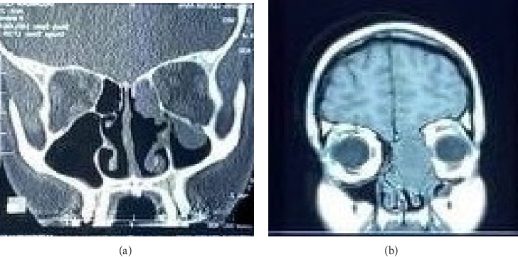
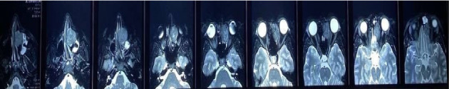
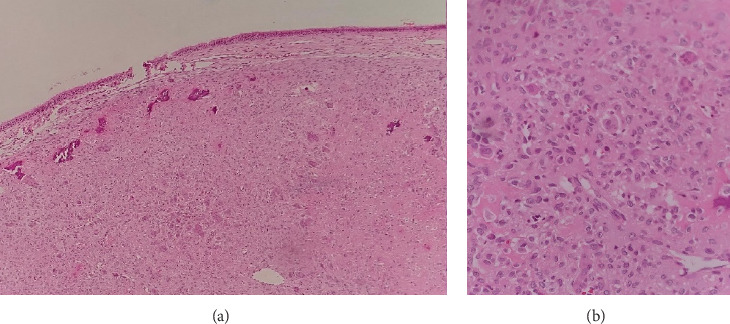
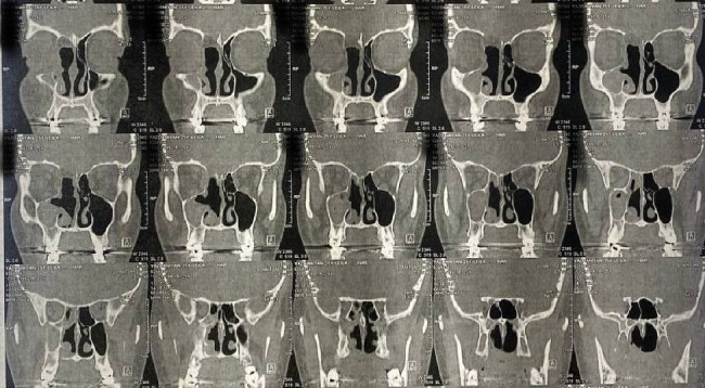
Background: Chondroblastoma is a rare and benign bone tumor originating from immature chondroblasts. Chondroblastoma typically affects long bones; however, it can also occur in the skull, especially the temporal bone. The anterior skull base is a rare location for this tumor, with only two reported cases. Case Presentation: A 26-year-old woman presented with epiphora in her right eye and progressive proptosis on the same side. She had a previous biopsy that confirmed the presence of a giant cell tumor of the bone and had undergone an unsuccessful endoscopic surgery. A comprehensive endoscopic procedure subsequently revealed a cartilage-producing neoplasm consistent with chondroblastoma. Conclusion: We presented a successful case of surgical resection of a chondroblastoma in the anterior skull base. Additionally, we reviewed the existing literature and previously documented cases.
Copyright © 2025 Mohsen Fazli et al. Case Reports in Otolaryngology published by John Wiley & Sons Ltd.
Conflict of interest statement
The authors declare no conflicts of interest.
Figures
Similar articles
-
Anterior Approach Total Ankle Arthroplasty with Patient-Specific Cut Guides.JBJS Essent Surg Tech. 2025 Aug 15;15(3):e23.00027. doi: 10.2106/JBJS.ST.23.00027. eCollection 2025 Jul-Sep. JBJS Essent Surg Tech. 2025. PMID: 40821726 Free PMC article.
-
Prescription of Controlled Substances: Benefits and Risks.2025 Jul 6. In: StatPearls [Internet]. Treasure Island (FL): StatPearls Publishing; 2025 Jan–. 2025 Jul 6. In: StatPearls [Internet]. Treasure Island (FL): StatPearls Publishing; 2025 Jan–. PMID: 30726003 Free Books & Documents.
-
Morphological, functional and neurological outcomes of craniectomy versus cranial vault remodeling for isolated nonsyndromic synostosis of the sagittal suture: a systematic review.JBI Database System Rev Implement Rep. 2015 Sep;13(9):309-68. doi: 10.11124/jbisrir-2015-2470. JBI Database System Rev Implement Rep. 2015. PMID: 26470674
-
[Recurrence of chondroblastoma in the distal femur: a clinical case report].Acta Ortop Mex. 2025 May-Jun;39(3):180-186. Acta Ortop Mex. 2025. PMID: 40645790 Spanish.
-
A Rare Case of Osteoblastic Osteosarcoma of the Temporal Bone and Comprehensive Review of the Literature.Otol Neurotol. 2025 Jun 1;46(5):e198-e201. doi: 10.1097/MAO.0000000000004481. Epub 2025 Feb 24. Otol Neurotol. 2025. PMID: 40062377
References
Publication types
LinkOut - more resources
Full Text Sources
