Case Reports
. 2025 Jul 2;10(3):owaf016.
doi: 10.1093/fsr/owaf016. eCollection 2025 Sep.

E-dentification, the use of teledentistry for remote personal forensic identification in forensic odontology: a Queensland experience

Affiliations
Case Reports

E-dentification, the use of teledentistry for remote personal forensic identification in forensic odontology: a Queensland experience

Neil Evans et al. Forensic Sci Res. .

Abstract

Dental comparison is recognized by the International Criminal Police Organization as one of three primary forensic identification techniques that can provide conclusive findings. Queensland is a large Australian state with a centralized forensic odontology service located at Queensland Health's Coronial and Public Health Sciences (CPHS) in Brisbane, which sits in the state's South-Eastern corner. Almost half of the Queensland population is located outside of Brisbane, and the distance to regional centres can be very large. Transporting forensic dental personnel and their equipment to these regional centres to undertake identification and examination procedures can be both expensive and time-consuming, depriving CPHS of service for the period of absence. The acquisition of post-mortem computed tomography (PMCT) data locally in regional centres with remote access electronically from CPHS in Brisbane has the potential to alleviate these issues in many cases. Forensic radiographers at CPHS work with forensic odontologists to produce multi-planar reformat images from PMCT data, which simulate common dental radiographs such as orthopantomogram, bitewing, and periapical views. Additional images, such as three-dimensional (3D) reconstructions of the teeth and jaws, can also be produced and viewed from various angles. These multi-planar reformat and 3D images can be compared with antemortem (AM) radiographic images and dental records of a missing person sourced from public or private dental surgeries, public hospitals, or private radiology practices. Comparisons can be made not only with AM traditional dental radiographs but also with images and reconstructions produced from AM dental cone-beam computed tomography or medical computed tomography data. The authors term this remote dental identification "e-dentification". While e-dentification offers numerous advantages, there are several limitations to its use, including access to the necessary equipment, the consistent acquisition of high-resolution PMCT data, and artefacts, including those due to metal restorations, that may be present in computed tomography images. We present four cases to illustrate and discuss e-dentification.

Keywords: computed tomography; forensic identification; forensic odontology; radiology; teledentistry.


Figures

Figure 1
Figure 1
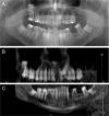
(A) Antemortem (AM) orthopantomogram (OPG) of a missing elderly male of interest in Case 1. An inverted impaction of tooth 18 as well as impactions of teeth 38, 48, and 23 are present, as well as retained deciduous teeth 63, 75, and 85. Teeth 27, 28, and 36 are missing, and multiple dental restorations are present. (B) Cropped multi-planar reformat (MPR) of the postmortem computed tomography (PMCT) data of the upper teeth of the deceased taken at the regional mortuary. The impactions of teeth 18 and 23 and the retained deciduous tooth 63 correspond to those in (A). The pattern of missing teeth is also concordant, although tooth 21 appears also to have been lost postmortem or postmortem (PM). (C) Cropped MPR of the PMCT data of the lower teeth of the deceased taken at the regional mortuary. The impactions of teeth 38 and 48 and the retained deciduous teeth 75 and 85 correspond to those in (A). The pattern of missing teeth is also concordant though tooth 34 also appears to have been lost perimortem or PM.
Figure 2
Figure 2
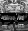
(A) Antemortem (AM) orthopantomogram (OPG) of the missing person of interest in Case 2. Root canal treatments (RCT) of teeth 14 and 24 are noted, as well as multiple dental restorations. A distinctive radiopacity is also noted in the left mandible. (B) Multi-planar reformat (MPR) image of the right side and anterior maxillary dentition. An RCT of tooth 14 is noted as well as a concordant pattern of dental restorations compared to (A). (C) Periapical-like view of teeth 25, 26, and 27. A concordant pattern of metallic restorations is noted compared to (A). (D) MPR image of the right side and anterior mandibular dentition. A concordant pattern of dental restorations, as well as concordant root and pulpal morphologies, is noted compared to (A). (E) MPR image of the left side mandibular dentition. A metallic restoration is present in tooth 37, and concordant root and pulpal morphologies are noted compared to (A). A distinctive radiopacity is noted distal to tooth 37, corresponding to that in (A).
Figure 3
Figure 3
(A) Antemortem (AM) orthopantomogram (OPG) of the missing person of interest in Case 3. Tooth 18 is mesioangularly impacted, and tooth 38 is absent. Two metallic occlusal restorations are noted in tooth 16. (B) Cropped multi-planar reformat (MPR) of the postmortem computed tomography (PMCT) data of the upper teeth of the deceased. Note the mesioangularly impacted tooth 18 and the appearance of the crown of tooth 16, although metallic restorations are not visible in this image. Tooth positions and root and pulpal morphologies are concordant with those in (A). (C) Cropped MPR of the PMCT data of the lower teeth of the deceased person. Note the correspondingly missing tooth 38 as well as the concordant tooth positions and root and pulpal morphologies. (D) PMCT three-dimensional (3D) reconstruction demonstrating metallic occlusal restorations of tooth 16. (E) PMCT 3D occlusal “bright” view demonstrating occlusal restorations of tooth 16.
Figure 4
Figure 4
Superimposition of the orthopantomogram (OPG) of a missing person over the postmortem computed tomography (PMCT) multi-planar reformat (MPR) of a deceased person indicates that both images originate from the same individual, establishing the identity. The impacted wisdom tooth shown in the OPG was recorded as having been subsequently extracted in the dental records of the missing person. (A–F) Opacities of 100%, 80%, 60%, 40%, 20%, and 0%, respectively.

References

    1. Geoscience Australia . Area of Australia – States and Territories. Australian Government. [cited 5 Oct 2022]. Available from: https://www.ga.gov.au/scientific-topics/national-location-information/di...
    1. Queensland Treasury. Queensland Population Counter. Queensland Government . [cited 5 Oct 2022]. Available from: https://www.qgso.qld.gov.au/statistics/theme/population/population-estim...
    1. Australian Bureau of Statistics . Regional Population 2021-2022 Financial Year. Australian Government; [cited 20 Jun 2023]. Available from: https://www.abs.gov.au/statistics/people/population/regional-population/...
    1. INTERPOL . INTERPOL Disaster Victim Identification Guide. INTERPOL, 2018. [cited 5 Oct 2022]. Available from: https://www.interpol.int/en/content/download/589/file/18Y1344%20E%20DVI_...
    1. Forrest A. Forensic odontology in DVI: current practice and recent advances. Forensic Sci Res. 2019;4:316–330. - PMC - PubMed

Publication types

LinkOut - more resources