Review
. 2025 Jul 31:15:1598552.
doi: 10.3389/fonc.2025.1598552. eCollection 2025.

Applications of 3D models in cholangiocarcinoma

Affiliations
Review

Applications of 3D models in cholangiocarcinoma

Agata Montagner et al. Front Oncol. .

Abstract

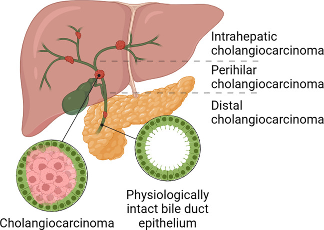
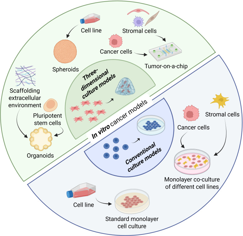
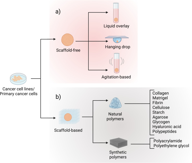
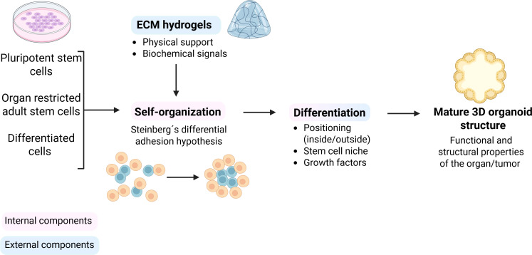
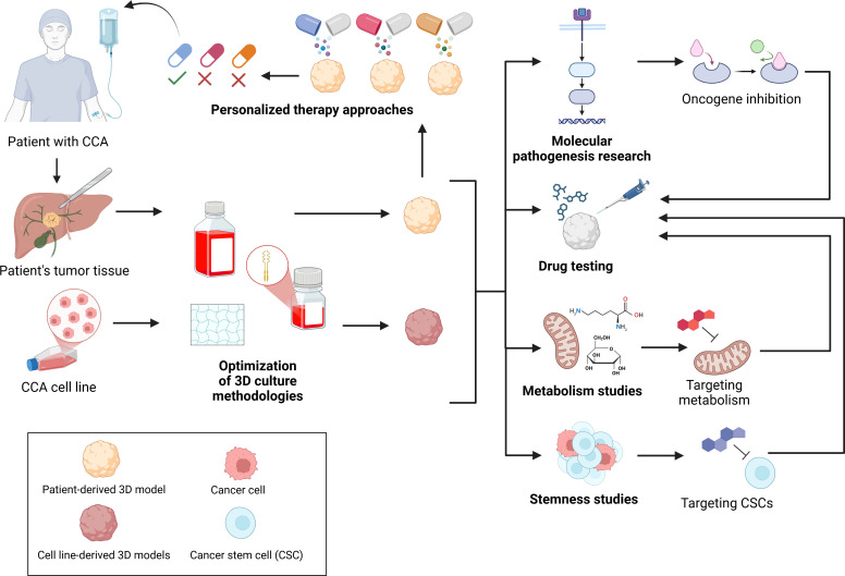
The prognosis for patients diagnosed with cholangiocarcinoma (CCA) is dismal, with an overall 5-year-mortality rate of 80%. Therapeutic approaches for this cancer are very limited and the only curative treatment is total surgical resection despite recent advancements in CCA research. However, only a minority of patients are eligible for surgery due to late-stage diagnosis. Therefore, there is an urgent need to gain a deeper understanding of CCA and to discover new treatments, which can be achieved by utilization and optimization of 3D tumor models. Traditional 2D cell culture is still undeniably important in cancer research, especially for the discovery of biomarkers and drug screening. However, classical 2D tumor models do not represent the tumor biology in its full complexity as they lack the vital interactions between cancer cells, angiogenesis, and tumor microenvironment. In recent years, 3D models, including spheroids, 3D co-culture systems, organoids, tumors-on-a-chip, and the in vivo chorioallantoic membrane (CAM) model, have been used for CCA research. These models enable the study of the tumor microenvironment, investigation of metastases, drug development and testing, cholangiocarcinogenesis and personalized therapy. This review summarizes the applications of the different 3D tumor models that have been used for the investigation of CCA. Moreover, the advantages and disadvantages of the different 3D tumor models are discussed, and suggestions for future research possibilities are described. By optimizing 3D models, the gap between basic research findings and clinical applications can be bridged, enabling the discovery of more effective therapies for CCA and other cancers.

Keywords: 3D (three-dimensional) models; CAM model; cancer; cholangiocarcinoma; in vitro cancer models; organoids; personalized medicine; tumor spheres.


Conflict of interest statement

The authors declare that the research was conducted in the absence of any commercial or financial relationships that could be construed as a potential conflict of interest.

Figures

Figure 1
Figure 1
Classification of CCAs according to anatomical location. Cholangiocarcinoma can be divided in intrahepatic, perihilar and distal cholangiocarcinoma. Created with Biorender.com.
Figure 2
Figure 2
Differences between two-dimensional and three-dimensional in vitro tumor models. One of the most diffused in vitro tumor models is the standard monolayer cell culture in which only one cell line is cultivated. In the co-culture monolayer method, more than one cell line is cultivated together but still in a two-dimensional system. Spheroids are simple free-floating cellular clusters. Organoids are complex 3D clusters in an arrangement that resembles the function and structure of the organ of origin. Tumors-on-a-chip are advanced models that couple cell culture with microfluidic devices. Created with Biorender.com.
Figure 3
Figure 3
Spheroid formation methodologies. Spheroids can be generated using (a) scaffold-free approaches, including liquid overlay, hanging drop, and agitation-based techniques, or (b) scaffold-based approaches employing either synthetic or natural polymers. Created with Biorender.com.
Figure 4
Figure 4
Organoids methodologies. 3D organoids can be generated from various cell sources, including pluripotent stem cells, organ restricted adult stem cells and differentiated cells. With the support of ECM-hydrogels providing physical structure and biochemical cues, cells undergo self-organization based on Steinberg’s differential adhesion hypothesis. This is followed by differentiation, influenced by positioning, stem cell niche and growth factors, ultimately leading to a mature 3D organoid structure that mimics the structural and functional features of the original organ or tumor. Created with Biorender.com.
Figure 5
Figure 5
Applications of spheroids and organoids in cholangiocarcinoma research. Tumor tissue from CCA patients or established CCA cell lines is used to generate 3D models through Optimized culture methodologies. These 3D models support studies regarding Molecular pathogenesis, Metabolism, Stemness, Drug Testing and Personalized therapy approaches. Created with Biorender.com.

Similar articles

References

    1. Sarcognato S, Sacchi D, Fassan M, Fabris L, Cadamuro M, Zanus G, et al. Cholangiocarcinoma. Pathologica. (2021) 113:158–69. doi: 10.32074/1591-951X-252, PMID: - DOI - PMC - PubMed
    1. Elvevi A, Laffusa A, Scaravaglio M, Rossi RE, Longarini R, Stagno AM, et al. Clinical treatment of cholangiocarcinoma: an updated comprehensive review. Ann Hepatol. (2022) 27:100737. doi: 10.1016/j.aohep.2022.100737, PMID: - DOI - PubMed
    1. Blechacz B, Komuta M, Roskams T, Gores GJ. Clinical diagnosis and staging of cholangiocarcinoma. Nat Rev Gastroenterol Hepatol. (2011) 8:512–22. doi: 10.1038/nrgastro.2011.131, PMID: - DOI - PMC - PubMed
    1. Cillo U, Fondevila C, Donadon M, Gringeri E, Mocchegiani F, Schlitt HJ, et al. Surgery for cholangiocarcinoma. Liver Int. (2019) 39 Suppl 1:143–55. doi: 10.1111/liv.14089, PMID: - DOI - PMC - PubMed
    1. Esmail A, Badheeb M, Alnahar B, Almiqlash B, Sakr Y, Khasawneh B, et al. Cholangiocarcinoma: the current status of surgical options including liver transplantation. Cancers (Basel). (2024) 16. doi: 10.3390/cancers16111946, PMID: - DOI - PMC - PubMed

LinkOut - more resources