. 2025 Aug;169(8):e70192.
doi: 10.1111/jnc.70192.

Alterations of NMDAR Subunits in the Cerebrospinal Fluid Across Neurodegenerative and Immunological Disorders

Affiliations

Alterations of NMDAR Subunits in the Cerebrospinal Fluid Across Neurodegenerative and Immunological Disorders

Sergio Escamilla et al. J Neurochem. 2025 Aug.

Abstract

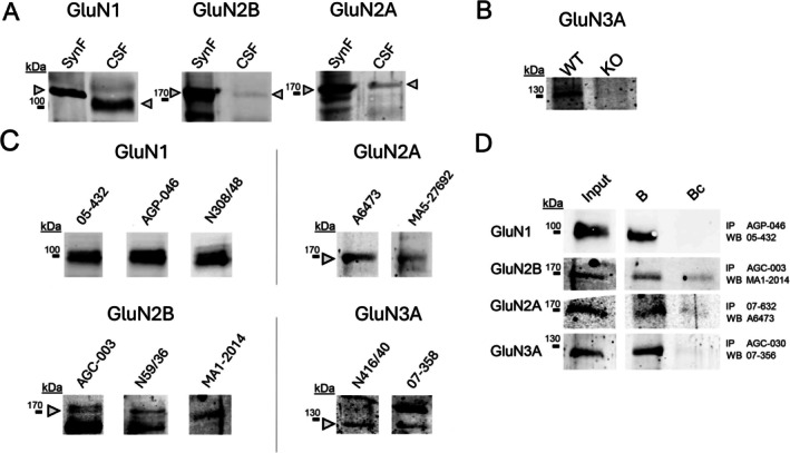
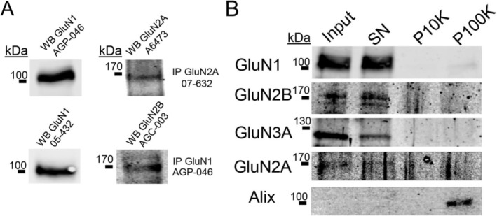
N-methyl-D-aspartate receptors (NMDARs) are glutamate-binding calcium channels that play a key role in brain function and have been linked to many neurological disorders. NMDARs are multi-pass membrane heterotetrameric complexes composed of two compulsory GluN1 subunits and two GluN2 (A-D) or GluN3 (A-B) subunits, from which GluN1, GluN2B, GluN2A, and GluN3A are widely expressed in the adult brain. This study assesses the presence of GluN1, GluN2B, GluN2A, and GluN3A in the cerebrospinal fluid (CSF) from healthy individuals, viral and autoimmune encephalitis, Huntington's disease (HD) and Alzheimer's disease (AD) patients. Samples were run in SDS-PAGE under reducing conditions and resolved with different anti-ectodomain and anti-C-terminal antibodies that combined with immunoprecipitation analyses, served to demonstrate the presence of full-length GluN1, GluN2A, GluN2B, and GluN3A in CSF. These NMDAR subunit complexes are not associated with extracellular vesicles. As a proof of concept of the identity of NMDAR subunits in the CSF, we demonstrated reduced levels of GluN1 in the CSF from patients with autoimmune encephalitis caused by anti-GluN1 antibodies compared with other causes of encephalitis; and showed a depletion of CSF GluN3A in a Grin3a knockout mouse model. Moreover, we observed higher GluN3A levels in CSF in both asymptomatic and symptomatic HD patients; while GluN2A levels were lower in CSF from AD patients. In conclusion, here we demonstrate the presence of NMDAR full-length subunits in CSF and that changes in NMDAR subunits balance could serve to identify alterations related to pathological conditions.

Keywords: Alzheimer's disease; CSF; GluN1; GluN2A; GluN2B; GluN3A; Huntington's disease; NMDAR; biomarker.


Conflict of interest statement

The authors have nothing to report.

The authors have stated explicitly that there are no conflicts of interest in connection with this article. APa received grant support from the Ministry of Health (MINSAL) and the Ministry of Education, Research and University (MIUR), from CARIPLO Foundation; personal compensation as a consultant/scientific advisory board member for Biogen, Lundbeck, Roche, Nutricia, General Healthcare (GE). APi received consultancy/speaker fees from Abbvie, Angelini, Bial, Lundbeck, Roche, and Zambon pharmaceuticals. H.Z. has served on scientific advisory boards and/or as a consultant for Abbvie, Acumen, Alector, Alzinova, ALZpath, Amylyx, Annexon, Apellis, Artery Therapeutics, AZTherapies, Cognito Therapeutics, CogRx, Denali, Eisai, LabCorp, Merry Life, Nervgen, Novo Nordisk, Optoceutics, Passage Bio, Pinteon Therapeutics, Prothena, Quanterix, Red Abbey Labs, reMYND, Roche, Samumed, Siemens Healthineers, Triplet Therapeutics, and Wave, has given lectures sponsored by Alzecure, BioArctic, Biogen, Cellectricon, Fujirebio, Lilly, Novo Nordisk, Roche, and WebMD, and is a co‐founder of Brain Biomarker Solutions in Gothenburg AB (BBS), which is a part of the GU Ventures Incubator Program (outside submitted work).

Figures

FIGURE 1
FIGURE 1
N‐methyl‐D‐aspartate receptor (NMDAR) subunits GluN2A, GluN2B, GluN1, and GluN3A are present in human cerebrospinal fluid (CSF). (A) Western blot of human synaptic membranes and CSF for GluN1 (antibody AGP‐046), GluN2B (antibody AGC‐003), and GluN2A (antibody A6473). (B) Western blot of CSF [pooling CSF obtained from 3 to 4 animals, and loading same volume (12 μL)] from wild‐type mouse (WT), and a mouse KO for Grin3a (GluN3A subunit, antibody 07‐356). (C) Combination of different antibodies detecting the same subunit, either GluN1 (C‐terminal: 05‐432; N‐terminal: AGP‐046 and N308/48), GluN2B (N‐terminal: AGC‐003 and N59/36; C‐terminal: MA1‐2014), GluN2A (C‐terminal: A6473 and MA5‐27692), or GluN3A (C‐terminal: 07‐356; N‐terminal: AGC‐030). (D) Immunoprecipitation (IP) of control CSF against the four different subunits using alternative antibodies to reveal the western blot (WB). Together with the input and the bound fraction (B), a control IP (Bc) was also revealed, corresponding to the incubation of the sample performed with an irrelevant IgG of the same animal species as the specific anti‐NMDAR antibody. Arrowheads point the immunoreactive band considered for further analysis. The inputs samples were resolved in the same gels but are shown separately to optimize contrast for defining discrete bands. The uncropped blots are available as Figure S2.
FIGURE 2
FIGURE 2
Co‐immunoprecipitation of N‐methyl‐D‐aspartate receptor (NMDAR) subunits and lack of the NMDAR immunoreactivities in extracellular vesicles. (A) The specific NMDAR subunit was immunoprecipitated (IP) from human cerebrospinal fluid (CSF) with the indicated antibody and assayed in immunoblots (WB) probed with an alternative antibody against the same subunit and also with an additional antibody against a different subunit, as indicated. (B) Western blot against the four different subunits using the following antibodies: GluN1 AGP‐046, GluN2B AGC‐003, GluN2A A6473, and GluN3A 06‐356. We used CSF as input and three different fractions of the extracellular vesicles (EVs) fractionation protocol (see Section 2 for details). Supernatant (SN) did not contain any EVs. Pellet P10K was obtained after centrifugation of 10 000 × g and contained apoptotic bodies and larger EVs. Pellet P100K was obtained after an ultracentrifugation of 100 000 × g and contained small EVs, as demonstrated by the presence of Alix, a canonical EV marker. The uncropped blot are available as Figure S3.
FIGURE 3
FIGURE 3
N‐methyl‐D‐aspartate receptor (NMDAR) subunit GluN1 levels in the cerebrospinal fluid (CSF) of anti‐NMDAR encephalitis patients. (A) Representative Western blots of GluN1 (antibody AGP‐046) subunit in samples from patients anti‐NMDAR (n = 4) encephalitis and encephalitis by other causes (n = 6; red is viral encephalitis, light blue is LG1 encephalitis and dark blue is anti‐Yo encephalitis). (B) GluN1 subunit was quantified. Data are expressed as percentages with respect to values from viral encephalitis samples. ROUT method identified no outliers. Exact P value obtained by t‐test is shown. Error bars represent SEM. The uncropped blot are available as Figure S4.
FIGURE 4
FIGURE 4
N‐methyl‐D‐aspartate receptor (NMDAR) subunit GluN3A levels in the cerebrospinal fluid (CSF) of Huntington's disease (HD) mutation carriers. (A) Representative Western blots of GluN3A (antibody 07‐356) and GluN1 (antibody AGP‐046) subunits in cases control (n = 9), cases of HD in the asymptomatic phase (H0; n = 10), and cases in the symptomatic phase (H1; n = 9). (B) Quantification of GluN3A subunit levels was normalized to the GluN1 subunit. An internal sample was used to normalize the values among different membranes. ROUT method identified 1 outlier from the control group and one from the H1 group, which were removed from the analysis. Data are expressed as percentages with respect to controls. Error bars represent SEM. Exact p values obtained by t‐test are shown. Given the exploratory nature of this study, we performed pairwise comparisons using independent t‐tests to specifically assess differences between C and each disease group (H0 and H1). The uncropped blot are available as Figure S5.
FIGURE 5
FIGURE 5
N‐methyl‐D‐aspartate receptor (NMDAR) subunits GluN2A and GluN2B levels in the cerebrospinal fluid (CSF) of patients with Alzheimer's disease (AD). (A) Representative western blots of GluN2A (antibody A6473), GluN2B (antibody AGC‐003), and GluN1 (antibody AGP‐046) subunits in human CSF from AD (n = 16) and non‐AD control cases (n = 17). An internal control sample was used to normalize among different membranes. (B) Quantification of GluN2A and (C) GluN2B subunit levels were normalized to the GluN1 subunit. ROUT method identified 3 outliers from the control group when measuring GluN2B, which were removed from the analysis. Data are expressed as percentages with respect to controls. Error bars represent SEM. Exact p value obtained by t‐test is shown. The uncropped blots are available as Figure S6.

Similar articles

References

    1. Bayés, À. , Collins M. O., Galtrey C. M., et al. 2014. “Human Post‐Mortem Synapse Proteome Integrity Screening for Proteomic Studies of Postsynaptic Complexes.” Molecular Brain 7: 88. 10.1186/s13041-014-0088-4. - DOI - PMC - PubMed
    1. Beaurain, M. , Salabert A. S., Payoux P., Gras E., and Talmont F.. 2024. “NMDA Receptors: Distribution, Role, and Insights Into Neuropsychiatric Disorders.” Pharmaceuticals (Basel) 17, no. 10: 1265. 10.3390/ph17101265. - DOI - PMC - PubMed
    1. Blennow, K. , and Zetterberg H.. 2018. “Biomarkers for Alzheimer‘s Disease: Current Status and Prospects for the Future.” Journal of Internal Medicine 284, no. 6: 643–663. 10.1111/joim.12816. - DOI - PubMed
    1. Crawley, O. , Conde‐Dusman M. J., and Pérez‐Otaño I.. 2022. “GluN3A NMDA Receptor Subunits: More Enigmatic Than Ever?” Journal of Physiology 600, no. 2: 261–276. 10.1113/JP280879. - DOI - PubMed
    1. Cuchillo‐Ibañez, I. , Lopez‐Font I., Boix‐Amorós A., et al. 2015. “Heteromers of Amyloid Precursor Protein in Cerebrospinal Fluid.” Molecular Neurodegeneration 10: 2. 10.1186/1750-1326-10-2. - DOI - PMC - PubMed

LinkOut - more resources