. 2025 Aug 11;53(15):gkaf763.
doi: 10.1093/nar/gkaf763.

Peripheral heterochromatin tethering is required for chromatin-based nuclear mechanical response

Affiliations

Peripheral heterochromatin tethering is required for chromatin-based nuclear mechanical response

Ali Göktuğ Attar et al. Nucleic Acids Res. .

Abstract

The cell nucleus is a mechanically responsive structure that governs how external forces affect chromosomes. Chromatin, particularly transcriptionally inactive heterochromatin, resists nuclear deformations through its mechanical response. However, chromatin also exhibits liquid-like properties, casting ambiguity on the physical mechanisms of chromatin-based nuclear elasticity. To determine how heterochromatin strengthens nuclear mechanical response, we performed polymer physics simulations of a nucleus model validated by micromechanical measurements and chromosome conformation capture data. The attachment of peripheral heterochromatin to the lamina is required to transmit forces directly to the chromatin and elicit its elastic response. Thus, increases in heterochromatin levels increase nuclear rigidity by increasing the linkages between chromatin and the lamina. Crosslinks within heterochromatin, such as HP1α proteins, can also stiffen nuclei, but only if chromatin is peripherally tethered. In contrast, heterochromatin affinity interactions that may drive liquid-liquid phase separation do not contribute to nuclear rigidity. When the nucleus is stretched, gel-like peripheral heterochromatin can bear stresses and deform, while the more fluid-like interior euchromatin is less perturbed. Thus, heterochromatin's internal structure and stiffness may regulate nuclear mechanics via peripheral attachment to the lamina, while also enabling nuclear mechanosensing of external forces and external measurement of the nucleus' internal architecture.


Conflict of interest statement

None declared.

Figures

Graphical Abstract
Graphical Abstract
Figure 1.
Figure 1.
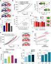
Heteropolymer simulation model recapitulates cell nuclear mechanical response. (A) Eight chromatid-like structures are relaxed into a phase-separated mixture of constitutive and facultative heterochromatin (blue and red) and euchromatin (green) within a polymeric lamina (gray). (B) Simulation snapshot with a schematic illustration of applied axial forces (top) and illustration of corresponding micromanipulation force spectroscopy experiment. (C) Representative force-strain curves measured in simulations of a chromatin-filled nucleus (gray) and an empty polymeric shell (i.e. only the lamina; black). Insets show simulation snapshots of the deformed morphologies; the empty shell inset also shows lamina buckles in a rotated view. (D) Fold change in short-extension spring constant from wildtype or typical simulation nuclei (turquoise or gray, respectively) to MNase-treated nuclei or empty polymeric shells (navy blue or black) regimes in experiments [28] and simulations.
Figure 2.
Figure 2.
Effects of heterochromatin microphase separation and internal structure on nuclear mechanical response. (A) Representative force-strain curves for nuclei with different fractions, f, of facultative heterochromatin, in nuclei with nonspecific affinity interactions between heterochromatin and the lamina. (B) The nuclear spring constant averaged over 10 replicates for various facultative heterochromatin levels, f, at different strains. (C) Nuclear spring constants computed from the slope of the force-strain curves at 30% strain. ** indicates p-values p < 0.01, while ns denotes not significant. (D) Nuclear spring constants at 30% strain as a function of the heterochromatin-lamina subunit attraction energy, EHL, and (E) the intra-heterochromatin subunit attraction energy, EHH. Inset images show representative snapshots of nuclear deformations and spatial organization of euchromatin (green), facultative heterochromatin (red), and constitutive heterochromatin (blue) contained within the nuclear lamina (gray). (F) Top left: schematic illustration of crosslinking within heterochromatin. Constitutive (blue) and facultative (red) heterochromatin may crosslink to other heterochromatin subunits of the same type. Bottom left: spring constants, knuc at 30% strain for different levels of crosslinking, set by the crosslink probability, Pc. Right: bar plot showing spring constants versus heterochromatin fraction, f, with Pc = 0.2 such that the total number of crosslinks increases with f. Data are shown as mean ± standard error of the mean (SEM). Statistical significance is assessed by one-way ANOVA followed by a post-hoc Tukey’s HSD test for pairwise comparisons. * denotes p < 0.05 and ** denotes p < 0.01.
Figure 3.
Figure 3.
Nuclear mechanical effects of heterochromatin tethering and changes in tethered heterochromatin organization. (A) Illustrations of specific tethering of heterochromatin to the lamina. (B) Left: Representative force-strain curves for nuclei with and without tethering of peripheral heterochromatin to the lamina. Right: Spring constants, knuc, as a function of strain. (C) Effective spring constants, knuc, at 30% strain, obtained from at least 10 independent force spectroscopy simulations. (D) Nuclear spring constant as a function of facultative heterochromatin tethering probability, PT. (E) Nuclear spring constant for various levels of heterochromatin, f, where f = 45% is the control case. The mean number of tethers, 〈NT〉, is either increased probabilistically (see the “Materials and methods” section) with f or held constant, as indicated by the numbers below each bar. Simulation snapshots are shown for increasing tethering number. (F) Left: Representative force-strain curves for nuclei with and without heterochromatin crosslinking in the presence or absence of facultative heterochromatin tethering. Right: Illustrations of these scenarios. (G) Schematic force-strain plot and illustration of expected scaling of force response as a function of relative stretched polymer length. Heterochromatin crosslinks act as constraints that decrease the length of stretchable polymer segments. (H) Fold change in effective nuclear spring constant from untethered and uncrosslinked scenario (purple) for nuclei with specific tethering and crosslinking only within different types of heterochromatin: constitutive (turquoise), facultative (yellow), and all (red). (I) Effective spring constants with different levels of crosslinked heterochromatin with lamina tethering. Left: knuc with tethered and crosslinked heterochromatin for different heterochromatin fractions, f, with crosslinking probability Pc = 0.2 such that the number of crosslinks increases with f. Right: knuc with tethered and crosslinked heterochromatin at fixed f, but with crosslinking levels determined by different crosslinking probabilities, Pc. Mean quantities of facultative and constitutive heterochromatin crosslinks, 〈NFHC〉 and 〈NCHC〉, respectively, are listed beneath each bar. Bars are mean ± SEM. Statistical significance is assessed by one-way ANOVA followed by a post-hoc Tukey’s HSD test for pairwise comparisons. * denotes p < 0.05, ** denotes p < 0.01, and *** denotes p < 0.001.
Figure 4.
Figure 4.
Chromatin-lamina tethering and crosslinking direct chromatin reorganization during nuclear deformation. (A) Simulated DamID of a 160 Mb region of chromosome 1 showing contact index of genomic sites before stretching (blue) and after 30% axial strain in nuclei with tethering and internal crosslinking of facultative heterochromatin (turquoise), tethering facultative heterochromatin without crosslinking (red), and untethered and uncrosslinked heterochromatin (gray). The contact index is 1 for chromatin subunits in contact with the lamina and 0 otherwise. The solid line shows a representative example, while shading indicates standard error. Right: 14 Mb snippet showing zoomed-in view of chromatin-lamina contacts for the region boxed on the left. (B) Top: Illustration of contact index. Bottom: Percentage of heterochromatin in contact with the lamina genome-wide, defined as distance r < 2.5σ, before stretching (navy blue bar) or after pulling to 30% strain in nuclei with and without heterochromatin tethering and/or crosslinking (gray, red, and turquoise). * indicates p < 0.05, ** indicates p < 0.01, and *** indicates p < 0.001. (C) Chromatin contact frequencies as a function of genomic distance for intra-heterochromatin contacts for the genomic distances of 4–40 Mb, before and after 30% nuclear strain for nuclei with both lamina tethering and heterochromatin crosslinking (left), tethering but no crosslinking (center), or neither tethering nor crosslinking (right). |Δrel| = |(Pafter(s) − Pbefore(s))/Pbefore(s)| indicates the absolute relative change in P(s) after deformation. (D) Chromatin contact frequencies as a function of genomic distance for intra-euchromatin contacts before and after nuclear deformation for different tethering and crosslinking scenarios. (E) Log-Log scaling plot showing the expected size of the internal deformation in chromatin as a function of strain for crosslinked (“gel”) and uncrosslinked (“unconstrained”) polymers. Illustrations show length scales over which deformations occur due to the presence or lack of constraining linkages. (F) Left: Absolute relative change, |Δrel|, in heterochromatic contacts as a function of genomic distance after deformation of nuclei with both lamina tethering and heterochromatin crosslinking (turquoise), tethering but no crosslinking (red), or neither tethering nor crosslinking (gray). Right: Bar plot showing the total absolute relative change, summed over all genomic distances. (G) Absolute relative change, |Δrel|, in euchromatic contacts as a function of genomic distance, with a bar plot showing the total change in contacts. Statistical significance is assessed by an unpaired, two-sided Student’s t-test.

Update of

Similar articles

Cited by

References

    1. Kalukula Y, Stephens AD, Lammerding J et al. Mechanics and functional consequences of nuclear deformations. Nat Rev Mol Cell Biol. 2022; 23:583–602. 10.1038/s41580-022-00480-z. - DOI - PMC - PubMed
    1. Miroshnikova YA, Wickström SA Mechanical forces in nuclear organization. Cold Spring Harb Perspect Biol. 2022; 14:a039685. 10.1101/cshperspect.a039685. - DOI - PMC - PubMed
    1. Nava MM, Miroshnikova YA, Biggs LC et al. Heterochromatin-driven nuclear softening protects the genome against mechanical stress-induced damage. Cell. 2020; 181:800–17. 10.1016/j.cell.2020.03.052. - DOI - PMC - PubMed
    1. Le HQ, Ghatak S, Yeung CYC et al. Mechanical regulation of transcription controls Polycomb-mediated gene silencing during lineage commitment. Nat Cell Biol. 2016; 18:864–75. 10.1038/ncb3387. - DOI - PubMed
    1. Golloshi R, Playter C, Freeman TF et al. Constricted migration is associated with stable 3D genome structure differences in cancer cells. EMBO Rep. 2022; 23:e52149. - PMC - PubMed

LinkOut - more resources