. 2025 Sep 10;16(9):e0198225.
doi: 10.1128/mbio.01982-25. Epub 2025 Aug 18.

c-di-GMP regulates bacterial NAD biosynthesis via targeting the transcriptional repressor NadR

Affiliations

c-di-GMP regulates bacterial NAD biosynthesis via targeting the transcriptional repressor NadR

Lianying Mao et al. mBio. .

Abstract

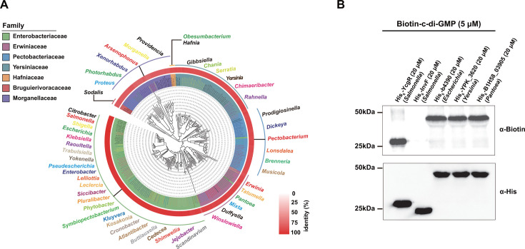
As a near-ubiquitous bacterial second messenger, cyclic di-GMP (c-di-GMP) regulates a multitude of important biological processes. The regulatory effects of c-di-GMP on bacterial physiological processes are mediated through its interaction with various effector molecules, including mRNA riboswitches and proteins. Although c-di-GMP effector proteins have been widely reported, yet unknown c-di-GMP effectors in bacteria wait to be discovered, and the physiological roles of this second messenger still remain to be explored. In a c-di-GMP/transcription factor binding screen, we identified NadR, a repressor of nicotinamide adenine dinucleotide (NAD) synthesis and salvage, as a c-di-GMP-responsive transcription factor in Salmonella enterica serovar Typhimurium. c-di-GMP was found to bind to NadR with high affinity. c-di-GMP binding inhibits the binding of NadR to its target DNA, thus upregulating the expression of NadR-repressed genes involved in NAD synthesis and salvage. c-di-GMP also stimulates the nicotinamide mononucleotide adenylyltransferase and ribosylnicotinamide kinase activities of NadR. As a result, elevated intracellular c-di-GMP levels lead to increased NAD synthesis and enhanced resistance to DNA damage in S. Typhimurium. NadR proteins from three other species belonging to Enterobacterales are capable of sensing c-di-GMP, suggesting that c-di-GMP-mediated modulation of intracellular NAD homeostasis is a conserved mechanism employed by members of Enterobacterales.

Importance: Cyclic di-GMP (c-di-GMP) functions as a highly versatile signaling molecule in bacteria, orchestrating diverse physiological processes critical for survival and adaptation. While nicotinamide adenine dinucleotide (NAD) plays pivotal roles in numerous cellular processes and functions, bacteria have been shown to modulate its biosynthetic and recycling pathways through a variety of regulatory mechanisms. However, a connection between c-di-GMP signaling and NAD metabolism in bacteria has never been revealed before. Here, we identify NadR, a transcriptional repressor of NAD synthesis and salvage, as a c-di-GMP effector, and show that c-di-GMP upregulates NAD biosynthesis by activating NadR-repressed genes, thus enhancing the defense of Salmonella against DNA damage. Our study reveals a previously unrecognized regulatory mechanism in bacterial NAD metabolism and expands the understanding of the physiological roles of c-di-GMP in bacteria.

Keywords: NAD biosynthesis; NAD metabolism; NadR; c-di-GMP.


Conflict of interest statement

The authors declare no conflict of interest.

Figures

Fig 1
Fig 1
Identification of NadR as a c-di-GMP receptor protein in S. Typhimurium. The UV-crosslinking assays detecting the binding of His6-NadR to biotinylated c-di-GMP. His6-YcgR was used as a positive control, and His6-InvF was used as a negative control. The reaction samples were separated by SDS-PAGE, transferred to nitrocellulose membrane, and probed with the anti-biotin antibody streptavidin horseradish peroxidase (top). The amount of each protein transferred to the membrane was also determined by western blot using the anti-His antibody (bottom). The blots presented are representative of three independent experiments with similar results.
Fig 2
Fig 2
c-di-GMP binds with high affinity and specificity to the NadR protein in S. Typhimurium. (A) The UV-crosslinking assays for binding specificity of His6-NadR to biotinylated c-di-GMP. Competition experiments were performed by the addition of unlabeled c-di-GMP, c-di-AMP, or cGMP simultaneously with biotinylated c-di-GMP to the reaction mixtures. (B) ITC assays for the interaction between NadR and c-di-GMP. The original titration data (the upper plot) and integrated heat measurements (the lower plot) are shown. The solid line in the lower plot represents the best fit to a one-site binding model of the interaction of NadR with c-di-GMP. (C) Schematic illustrating the domain organization of NadR. The HTH DNA-binding domain (residues 1–57), the NMN-AT domain (residues 58–227), and the RNam-K domain (residues 228–410) are indicated. (D) Predicted binding mode of c-di-GMP to the NMN-AT domain of NadR. (E) Schematic of the predicted contacts between c-di-GMP and NadR. Potential hydrogen bonds are indicated as green dashed lines. (F–J) ITC assays for the interactions of the NadR variants K72A (F), H80A (G), E197A (H), R212A (I), or H80A/R212A (J) with c-di-GMP. (A, B, and F–J) Data shown are representatives of three independent experiments with similar results. (B and F–J) Kd and complex stoichiometry (N) are presented as mean ± SD of three independent experiments.
Fig 3
Fig 3
c-di-GMP interferes with NadR binding to its target promoters but stimulates its NMN-AT and RNam-K activities. (A and B) EMSAs demonstrating specific binding of NadR to promoters of the nadB gene (A) and the nadA-pnuC operon (B). DNA fragments amplified from the coding regions of nadB (A) and nadA (B) were used as DNA negative controls, and BSA was used as the protein control. (C and D) EMSAs for NadR binding to the promoters of nadB (C) and nadA-pnuC (D) in the absence or presence of nucleotides. Nucleotides were added simultaneously with NadR to the reaction system. (E and F) EMSAs for the binding of NadR and its variants K72A, H80A, E179A, R212A, and H80A/R212A to the promoters of nadB (E) and nadA-pnuC (F) in the absence or presence of c-di-GMP. (G and H) c-di-GMP enhances the NMN-AT (G) and RNam-K (H) activities of NadR. (A–F) Gels shown are representative of three independent experiments with similar results. (G and H) Data are mean ± SD of three biological replicates. Two-sided, unpaired Student’s t-test was used for statistical analyses. **P < 0.01.
Fig 4
Fig 4
c-di-GMP, ATP, and NAD can competitively bind to NadR. (A) The UV-crosslinking assay showing binding of His6-NadR to biotinylated c-di-GMP in the absence or presence of unlabeled nucleotides or NAD. The unlabeled nucleotides or NAD were added simultaneously with biotinylated c-di-GMP to the reaction system containing His6-NadR. (B and C) EMSAs for NadR binding to the promoters of nadB (B) and nadA-pnuC (C) in the absence or presence of c-di-GMP and/or different concentrations of NAD. (D and E) EMSAs for the binding of NadR to the promoters of nadB (D) and nadA-pnuC (E) in the absence or presence of ATP, NAD, and/or different concentrations of c-di-GMP. (F and G) EMSAs for the binding of NadR to the promoters of nadB (F) and nadA-pnuC (G) in the absence or presence of ATP and c-di-GMP and/or different concentrations of NAD. (A–G) Blots and gels shown are representative of three independent experiments with similar results. (B–G) c-di-GMP, ATP, or NAD were added simultaneously with NadR to the reaction system.
Fig 5
Fig 5
Changes in intracellular c-di-GMP levels regulate the expression of genes involved in the de novo and salvage pathways of NAD biosynthesis via targeting NadR. (A) Intracellular c-di-GMP levels in the wild-type strain, the ΔnadR mutant, and the point mutant nadR(E179A) were modulated by overexpression of adrA or STM3611. (B–D) qRT-PCR analysis of the mRNA levels of nadB (B), nadA (C), and pnuC (D) in the wild-type strain, the ΔnadR mutant, the point mutant nadR (E179A), and their derivatives overexpressing adrA or STM3611. Expression levels were normalized to 16S rRNA and presented as values relative to that of the wild type without gene overexpression. (E and F) The promoter activities of the nadB gene (E) and the nadA-pnuC operon (F) in the wild type, ΔnadR, nadR(E179A), and their derivatives overexpressing adrA or STM3611. (G) Chromatin immunoprecipitation (ChIP)-qPCR quantifying binding of HA-NadR and HA-NadRE179A at the promoters of nadB and nadA-pnuC in the wild type, nadR(E179A), and their derivatives overexpressing adrA or STM3611. The ChIP-qPCR signals were normalized to their respective DNA inputs. (A–G) Data are mean ± SD of three biological replicates. Two-sided, unpaired Student’s t-test was used for statistical analyses. ns, not significant; **P < 0.01; ***P < 0.001; and ****P < 0.0001.
Fig 6
Fig 6
Changes in intracellular c-di-GMP levels modulate NAD biosynthesis via targeting NadR and enhance DNA damage resistance of S. Typhimurium. (A and B) Comparison of the intracellular level of NAD+ (A) and NADH (B) among the wild type, ΔnadR, nadR(E179A), and their derivatives overexpressing adrA or STM3611. (C) Survival rates of the wild type, ΔnadR, nadR(E179A), and their derivatives overexpressing adrA or STM3611 after exposure to UV irradiation. The data are shown as means ± SD of three biological replicates. Student’s t-test was used for statistical analyses. ns, not significant; *P < 0.05; **P < 0.01; ***P < 0.001; and ****P < 0.0001.
Fig 7
Fig 7
NadR homologs within the order Enterobacterales also bind c-di-GMP. (A) Phylogenetic analysis of the NadR family in the order Enterobacterales. NadR homologs were searched by BLASTP against the NCBI non-redundant protein database, and members of the order Enterobacterales were used for phylogenetic analysis (Data S1). The heat map represents the sequence identity between the NadR protein in S. Typhimurium and its homologs. (B) The UV-crosslinking assay showing the binding of three NadR proteins, His6-b4390, His6-YPK_3620, and His6-B1H58_03905, from E. coli, Y. pseudotuberculosis, and P. alhagi, respectively, to biotinylated c-di-GMP. The blots shown are representative of three independent experiments with similar results.

References

    1. Jenal U, Reinders A, Lori C. 2017. Cyclic di-GMP: second messenger extraordinaire. Nat Rev Microbiol 15:271–284. doi: 10.1038/nrmicro.2016.190 - DOI - PubMed
    1. Khan F, Jeong GJ, Tabassum N, Kim YM. 2023. Functional diversity of c-di-GMP receptors in prokaryotic and eukaryotic systems. Cell Commun Signal 21:259. doi: 10.1186/s12964-023-01263-5 - DOI - PMC - PubMed
    1. Römling U, Galperin MY, Gomelsky M. 2013. Cyclic di-GMP: the first 25 years of a universal bacterial second messenger. Microbiol Mol Biol Rev 77:1–52. doi: 10.1128/MMBR.00043-12 - DOI - PMC - PubMed
    1. Li S, Sun H, Li J, Zhao Y, Wang R, Xu L, Duan C, Li J, Wang Z, Liu Q, Wang Y, Ouyang S, Shen X, Zhang L. 2022. Autoinducer-2 and bile salts induce c-di-GMP synthesis to repress the T3SS via a T3SS chaperone. Nat Commun 13:6684. doi: 10.1038/s41467-022-34607-9 - DOI - PMC - PubMed
    1. Li S, Liu Q, Duan C, Li J, Sun H, Xu L, Yang Q, Wang Y, Shen X, Zhang L. 2023. C-di-GMP inhibits the DNA binding activity of H-NS in Salmonella. Nat Commun 14:7505. doi: 10.1038/s41467-023-43442-5 - DOI - PMC - PubMed

MeSH terms

LinkOut - more resources