. 2025 Aug;21(8):e70550.
doi: 10.1002/alz.70550.

A brain-derived tau oligomer polymorph is associated with cognitive resilience to Alzheimer's disease

Affiliations

A brain-derived tau oligomer polymorph is associated with cognitive resilience to Alzheimer's disease

Michela Marcatti et al. Alzheimers Dement. 2025 Aug.

Abstract

Introduction: Misfolded tau can assemble into oligomers that adopt distinct conformations, referred to as polymorphs, each with unique biochemical and pathological properties. These tau polymorphs are thought to influence disease progression in Alzheimer's disease (AD) and related disorders. Interestingly, some individuals with AD pathology remain cognitively intact (non-demented with Alzheimer's neuropathology [NDAN]), suggesting potential differences in tau polymorph profiles.

Methods: Brain-derived tau oligomers (BDTOs) were isolated from post mortem hippocampi of AD and NDAN individuals. Their biophysical, biochemical, and functional properties were assessed via protease digestion, immunocharacterization, atomic force microscopy, tau seeding in biosensor cells, hippocampal slice electrophysiology, and SH-SY5Y toxicity assays.

Results: NDAN-BDTOs exhibited protease resistance, different conformational profiles, formed larger aggregates, preserved synaptic function, and reduced neuronal toxicity compared to AD-BDTOs.

Discussion: The data suggests that a structurally stable yet less toxic tau polymorph in NDAN may underlie cognitive resilience, supporting the therapeutic relevance of targeting specific tau polymorphs.

Highlights: Non-demented with Alzheimer's neuropathology (NDAN) brain-derived tau oligomers (BDTOs) exhibit distinct, more stable structural polymorphs compared to those from Alzheimer's disease (AD). NDAN-BDTOs are larger, more protease resistant, and less toxic in cell and hippocampal slice models. Both NDAN- and AD-BDTOs seed tau aggregation, but NDAN-BDTOs promote formation of less toxic assemblies. Structural stability of NDAN-BDTOs may contribute to cognitive resilience despite AD pathology. Mimicking non-toxic tau polymorphs could represent a novel therapeutic strategy for AD.

Keywords: Alzheimer's disease; brain‐derived tau oligomers; cognitive resilience; non‐demented with Alzheimer's neuropathology; tau polymorphs.


Conflict of interest statement

The authors declare no conflicts of interest. Author disclosures are available in the supporting information.

Figures

FIGURE 1
FIGURE 1
Schematic overview of experimental design. BDTOs were isolated from AD and NDAN brain tissues and characterized using biochemical, biophysical, and biological assays. This integrative approach revealed the presence of two different tau polymorphs in AD and NDAN individuals. AD, Alzheimer's disease; BDTO, brain‐derived tau oligomer; IP, immunoprecipitation; LDH, L‐lactate dehydrogenase; NDAN, non‐demented with Alzheimer's neuropathology.
FIGURE 2
FIGURE 2
Validation of isolation and immunoreactivity profiles of BDTOs from AD and NDAN individuals. A, Immunoprecipitation of BDTOs from AD and NDAN individuals. Representative western blot showing tau immunoreactivity detected with a recombinant monoclonal anti‐tau antibody in input (I), unbound (UB), immunoglobulin G isotype control (IgG), and immunoprecipitated eluates (IPE1 and IPE2) from pooled hippocampal homogenates of age‐matched AD and NDAN individuals. BDTOs were isolated using the in‐house anti‐oligomeric tau antibody T18. Tau enrichment in IPE1 and IPE2 confirms successful and consistent immunoprecipitation of BDTOs from both groups. B, Immunoreactivity profiles of BDTOs from AD and NDAN individuals. Representative western blot analyses of BDTOs from AD and NDAN individuals (IPE1 and IPE2), probed with antibodies recognizing total tau (recombinant monoclonal anti‐tau) and oligomeric tau (T22 and TOMA2). Each blot includes a standard curve of recombinant tau oligomers (rTauO) of known concentrations. NDAN‐BDTOs displayed reduced reactivity to the monoclonal anti‐tau antibody, similar reactivity to T22, and increased reactivity to TOMA2 compared to AD‐BDTOs. These patterns were consistent across both IPE1 and IPE2 fractions, with IPE2 showing overall lower levels of immunoreactivity. AD, Alzheimer's disease; BDTO, brain‐derived tau oligomer; IP, immunoprecipitation; NDAN, non‐demented with Alzheimer's neuropathology.
FIGURE 3
FIGURE 3
Morphological characterization of BDTOs from AD and NDAN individuals. A, Representative AFM images of BDTOs immunoprecipitated from AD and NDAN hippocampal tissue. B, Distribution histograms and (C) quantification of BDTOs’ diameter shows a significant increase in NDAN compared to AD, represented as fold change relative to AD. D–E, Similar analysis for BDTOs’ height confirms greater vertical dimensions in NDAN‐derived BDTOs. Data indicate that NDAN BDTOs are larger in both diameter and height than their AD counterparts. Data are represented as mean ± standard deviation; biological replicates n = 3; P* < 0.05 and **P < 0.005; two‐tailed t test. AD, Alzheimer's disease; AFM, atomic force microscopy; BDTO, brain‐derived tau oligomer; IP, immunoprecipitation; NDAN, non‐demented with Alzheimer's neuropathology.
FIGURE 4
FIGURE 4
Proteolytic profiling of BDTOs from NDAN and AD individuals. Representative western blot of BDTOs immunoprecipitated from AD and NDAN hippocampal tissue exposed to increasing concentrations of proteinase K (0, 0.5, 1, and 2.5 µg/mL) for 1 hour at 37°C. Blots were probed with Tau5 (left) and Tau13 (right) antibodies to assess proteolytic profiles. NDAN‐BDTO showed greater resistance to degradation compared to AD‐BDTO, suggesting the presence of structurally distinct and more protease‐resistant tau conformers in NDAN. AD, Alzheimer's disease; BDTO, brain‐derived tau oligomer; NDAN, non‐demented with Alzheimer's neuropathology; PK, proteinase K.
FIGURE 5
FIGURE 5
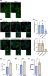
Seeding activity of BDTOs from AD and NDAN. A, Representative FRET images of untreated TauRD(P301S) biosensor cells (CTR) and cells treated with liposome (vehicle), showing no detectable tau aggregation. B, FRET images of biosensor cells treated with 0.25 µM BDTOs from AD and NDAN individuals for 24 hours. Both AD and NDAN BDTOs induced tau aggregation, as evidenced by fluorescent tau aggregates. Cells treated with BDTOs pre‐incubated with an IgG isotype control (middle panel) or BDTOs pre‐incubated with T22 antibody (right panel) are shown. C, Quantification of tau aggregates after BDTOs treatment, comparing the effects of the T22 antibody on seeding activity; data represent the mean ± SD; P* < 0.05; One‐way analysis of variance followed by Dunnett post hoc test. D–F, Morphometric analysis of tau aggregates induced by BDTOs. Volume (D), area (E), and mean fluorescent intensity (MFI; F) were measured for each BDTO‐treated group. Data represent the mean ± SD; biological replicates n = 3; P* < 0.05 and **P < 0.005; two‐tailed t test. AD, Alzheimer's disease; BDTO, brain‐derived tau oligomer; FRET, fluorescence resonance energy transfer; IgG, immunoglobulin G; NDAN, non‐demented with Alzheimer's neuropathology; SD, standard deviation.
FIGURE 6
FIGURE 6
Electrophysiological analysis of synaptic function in hippocampal slices treated with BDTOs from AD and NDAN. A, Representative trace before (baseline) and after (post‐) high frequency stimulation (HFS); (B) Percentage of the EPSP slope. HFS was performed at the end of the 10th minute; (C) Average of the last 10 minutes of the EPSP slope showed that BDTOs from NDAN individuals are less toxic than those from AD; (D) Representative traces of pair‐pulse stimulation with 50 ms interstimulus interval (ISI); (E) paired‐pulse stimulation (PPS) analysis showing impaired paired‐pulse facilitation in slices treated with AD‐BDTOs, indicating dysfunction in short‐term plasticity. Minimum of three different mice each group, each point represents data from a single half‐brain slice (ACSF n = 7; AD‐BDTOs n = 12; NDAN‐BDTOs n = 12, see the Statistical analysis section for criteria of exclusion). Data represent the mean ± standard error of the mean; P* < 0.05, **P < 0.005, ***P < 0.001; one‐way analysis of variance followed by a Tukey post hoc test. ACSF, artificial cerebrospinal fluid; AD, Alzheimer's disease; BDTO, brain‐derived tau oligomer; EPSP, excitatory post‐synaptic potential; HFS, high‐frequency stimulation; IgG, immunoglobulin G; NDAN, non‐demented with Alzheimer's neuropathology; SD, standard deviation.
FIGURE 7
FIGURE 7
Cytotoxicity analysis of BDTOs from AD and NDAN individuals. A, LDH release from SH‐SY5Y cells treated for 4 hours with increasing concentrations (50, 100, and 200 nM) of BDTOs from AD and NDAN individuals to determine the optimal concentration for the assay. Data represent the mean ± SD; biological replicates, n = 3; *P < 0.05; one‐way ANOVA followed by a Tukey post hoc test. B, SH‐SY5Y cells were treated with the selected concentration (50 nM) of BDTOs, BDTOs pre‐incubated with IgG isotype control, or BDTOs pre‐incubated with T22 antibody to assess the specificity of the cytotoxic effect. Data represent the mean ± SD; each dot represents a biological replicate (CTR, n = 5; AD, n = 7; AD+IgG, n = 4; AD+T22, n = 6; NDAN, n = 6; NDAN+IgG, n = 4; NDAN+T22, n = 4); **P < 0.005, ***P < 0.001; one‐way ANOVA followed by a Tukey post hoc test. C, Representative plots showing the gating strategy for cytofluorimetric analysis of propidium iodide (PI) incorporation. The upper left panel shows bead‐based gating; the upper right panel shows gating for SH‐SY5Y cells. Lower panels display the percentage of PI‐positive (PI⁺) cells for control, AD‐treated, and NDAN‐treated groups based on these gates. D, Histograms of mean fluorescence intensity (MFI) of PI⁺ SH‐SY5Y cells for all treatment conditions, generated from combined data across all biological replicates (n) to illustrate overall distribution patterns. E, Quantification of MFI values shown in (D), expressed as fold change relative to untreated control cells. Data represent the mean ± SD; biological replicates, n = 5; *P < 0.05, **P < 0.005, ***P < 0.001; one‐way ANOVA followed by a Tukey post hoc test. AD, Alzheimer's disease; ANOVA, analysis of variance; BDTO, brain‐derived tau oligomer; IgG, immunoglobulin G; LDH, L‐lactate dehydrogenase; NDAN, non‐demented with Alzheimer's neuropathology; SD, standard deviation.

References

    1. Fabiani C, Antollini SS. Alzheimer's disease as a membrane disorder: spatial cross‐talk among beta‐amyloid peptides, nicotinic acetylcholine receptors and lipid rafts. Front Cell Neurosci. 2019;13:309. doi: 10.3389/fncel.2019.00309 - DOI - PMC - PubMed
    1. Tiwari S, Atluri V, Kaushik A, Yndart A, Nair M. Alzheimer's disease: pathogenesis, diagnostics, and therapeutics. Int J Nanomedicine. 2019;14:5541‐5554. doi: 10.2147/IJN.S200490 - DOI - PMC - PubMed
    1. Spires‐Jones TL, Hyman BT. The intersection of amyloid beta and tau at synapses in Alzheimer's Disease. Neuron. 2014;82(4):756‐771. doi: 10.1016/j.neuron.2014.05.004 - DOI - PMC - PubMed
    1. Ashraf GM, Greig NH, Khan TA, et al. Protein Misfolding and Aggregation in Alzheimer's Disease and Type 2 Diabetes Mellitus HHS Public Access . Vol 13.; 2014. - PMC - PubMed
    1. John A, Reddy PH. Synaptic basis of Alzheimer's disease: focus on synaptic amyloid beta, P‐tau and mitochondria. Ageing Res Rev. 2021;65:101208. doi: 10.1016/j.arr.2020.101208 - DOI - PMC - PubMed