. 2025 Sep 10;16(9):e0100825.
doi: 10.1128/mbio.01008-25. Epub 2025 Aug 18.

Encephalomyocarditis virus protein 2B* interacts with 14-3-3 proteins through a phosphorylated C-terminal binding motif

Affiliations

Encephalomyocarditis virus protein 2B* interacts with 14-3-3 proteins through a phosphorylated C-terminal binding motif

Samantha K Nguyen et al. mBio. .

Abstract

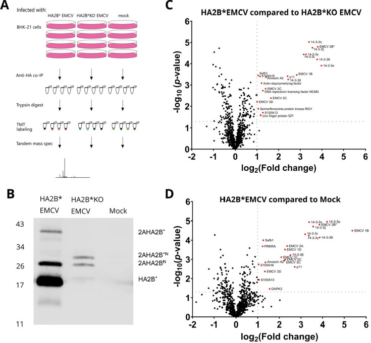
Encephalomyocarditis virus (EMCV) has been an important model RNA virus for decades. Although most of the EMCV proteins are obtained via proteolytic cleavage of a long polyprotein, 2B* is expressed from a short overlapping open reading frame via an unusual protein-stimulated temporally dependent ribosomal frameshifting mechanism. Mutant viruses that are unable to express 2B* have a small plaque phenotype due to delayed onset and inefficient progression of multiple lytic cell death pathways. However, the mechanism by which 2B* promotes these pathways has not yet been characterized. By tagging 2B* within the viral genome, we identified putative interaction partners of 2B* and showed that 2B* binds all seven members of the 14-3-3 protein family during virus infection. Binding is entirely dependent on a mode III motif containing a phosphoserine (RRNpSS) at the 2B* C terminus. This may impede other functions of the 14-3-3 proteins, including their role in promoting antiviral signaling. Ablating the 2B*:14-3-3 interaction had no effect upon plaque size, indicating that the function of this interaction is unrelated to the role of 2B* in promoting lytic cell death. This work expands our knowledge of the protein complement of this important model virus and the binding repertoire and specificity of host 14-3-3 proteins.IMPORTANCEEncephalomyocarditis virus (EMCV) infects a range of species, causing economically important reproductive disorders in pigs and encephalitis and myocarditis in rodents. Due to its wide host range, it is an important model pathogen for investigating virus-host interactions. EMCV expresses an accessory protein, 2B*, from an overlapping open reading frame via an unusual ribosomal frameshifting mechanism. Although the frameshifting mechanism has been established, the function of the 2B* protein had not been explored until recently. Here, we determined the host proteins to which 2B* binds and found that it specifically binds to all members of the 14-3-3 protein family, which, among other roles, contribute to the innate immune response to viral infection in mammalian cells. The interaction requires a specific stretch of amino acids at the end of 2B*. Binding to 2B* may reduce the opportunities for these 14-3-3 proteins to bind to host proteins and perform their usual roles; therefore, by interacting with the 14-3-3 proteins, 2B* may affect multiple host cell functions, including immune response activation.

Keywords: 14-3-3 proteins; 2B*; EMCV; cardiovirus; innate immunity; interferon beta; picornavirus; proteomics; ribosome frameshifting.


Conflict of interest statement

The authors declare no conflict of interest.

Figures

Fig 1
Fig 1
2B*KO EMCV mutations do not impact viral frameshifting or replication. (A) Schematic representation of the dual luciferase constructs. Renilla luciferase is produced by every translating ribosome. However, in the WT sequence, firefly luciferase is only produced when frameshifting does not occur. This enables quantitative measurement of the percentage of ribosomes in each reading frame for each mutant construct. (B) BHK-21 cells were transiently co-transfected with a WT EMCV 2A expression construct (pCAGG-2A) along with a dual luciferase plasmid containing 105 nt of the EMCV genome (WT pSGDLuc) or mutants thereof (2B*KO pSGDLuc, SS-SL pSGDLuc, or −1 SS-SL pSGDLuc) at various ratios. The SS-SL cassette contains mutations in both the slippery sequence and the stem-loop, which collectively ablate PRF. The −1 SS-SL cassette contains the SS-SL mutations, as well as an additional single nucleotide insertion, ensuring all ribosomes enter the second ORF. This construct therefore mimics 100% frameshifting. The 2B*KO cassette contains the premature termination codons, used elsewhere in this manuscript, designed to ablate 2B* expression while leaving PRF rates unaffected. At 24 h post-transfection, cells were frozen in 1× passive lysis buffer, and both renilla and firefly luciferase activities were measured. Samples were normalized to the luciferase values for the same pSGDLuc construct co-transfected with pCAGG-2Amut. The percentages of ribosomes in the 0 or −1 reading frames for each ratio of pCAGG-2A:pSGDLuc were calculated. Data shown are the mean ± SD of three biological repeats, each using triplicate wells. Statistical analysis (Student’s t-test): ns, not significant; *P ≤ 0.05; and *P ≤ 0.01. (C) A confluent monolayer of BHK-21 cells was infected with GFP WT EMCV or GFP 2B*KO EMCV at an MOI of 0.01. An equivalent volume of cell lysate from GFP GNN EMCV-transfected cells was also included as an additional control. GCU was analyzed using the Incucyte live-cell imaging suite (Sartorius) and normalized to the respective GCU at 5.5 hpi. Data shown are the mean ± SD of three biological repeats, each using triplicate wells. Statistical analysis (Student’s t-test of the indicated virus compared to GFP WT EMCV): ns, not significant; *P ≤ 0.05.
Fig 2
Fig 2
Characterization of HA2B* EMCV. (A) Schematic representation of the EMCV viral genome with sequence encoding the HA tag inserted to tag 2B*. (B) BHK-21 cells were infected with WT EMCV, 2B*KO EMCV, HA2B* EMCV, or HA2B*KO EMCV at an MOI of 5.0. Cells were harvested at the indicated time points and frozen in complete RIPA buffer. Lysates were subjected to SDS-PAGE and immunoblotting. The membrane was cut at the 25 kDa marker on the ladder to allow the same membrane to be probed by all four antibodies. The molecular mass scale (kDa) is indicated at the left, and antibodies used are labeled at the right. (C) Images of plaques formed by the indicated viruses, grown in BSR cells. Images are representative of three independent biological repeats. (D) Size distribution of plaques formed by WT EMCV, HA2B* EMCV, and their respective 2B*KO mutants. Distributions shown are based on area measurements of 30 plaques, sampled from three biological repeats. Horizontal lines represent the median (dashed) and upper and lower quartiles (dotted). Statistical analysis (ratio-paired t-test): ****P ≤ 0.0001. (E) BHK-21 cells were infected with the indicated viruses at an MOI of 3. At the indicated time points post-infection, released and intracellular virus was harvested, and the titer of each was determined by plaque assay. Total infectious virus (PFU/mL) was calculated for each virus at each time point. Data represent the means ± SEM of two independent biological repeats.
Fig 3
Fig 3
Putative interaction partners of 2B* include the entire family of 14-3-3 proteins. (A) Schematic diagram of the TMT experiment. BHK-21 cells were infected with either HA2B* EMCV or HA2B*KO EMCV, or mock infected. At 7 hpi, HA2B* and any interaction partners were purified from the lysates by anti-HA co-immunoprecipitation; 87.5% of each sample was then used for trypsin digestion, TMT labeling, and analysis by mass spectrometry. (B) The remaining 12.5% of each co-immunoprecipitated sample was probed for HA-tagged proteins by western blot using an anti-HA antibody. The molecular mass scale (kDa) is indicated at left, and HA-tagged proteins are labeled at right. (C and D) Two-sample t-tests were used to compare protein enrichment in the HA2B* EMCV-infected samples relative to HA2B*KO EMCV-infected samples (panel C) and mock-infected samples (panel D). Candidate interaction partners were defined as those with −log10(P-value) greater than 1.3 (i.e., P-value > 0.05) and log2(fold change) > 1 (i.e., fold change greater than 2) and are indicated with red dots and labeled. Note that, due to the presence of background noise, the infected versus mock fold changes for virus proteins are not infinite.
Fig 4
Fig 4
2B* binds to the entire family of 14-3-3 proteins via a C-terminal RRNSS sequence. (A) BHK-21 cells were co-transfected with equal amounts of pCAGG-HA2B* and the specified N-terminally FLAG-tagged 14-3-3 encoding plasmid (pCAGG-FLAG 14-3-3x), 24 h prior to immunoprecipitation via the FLAG epitope. Samples were then subjected to SDS-PAGE and immunoblotting using the indicated antibodies. Data shown are representative of two independent biological repeats. (B) BHK-21 cells were co-transfected with both a plasmid encoding either an N-terminally FLAG-tagged 14-3-3 protein or tubulin β chain and another encoding either HAΔRRNSS2B* or HA2B*, as indicated. Cell lysates were immunoprecipitated with anti-FLAG antibody and subjected to SDS-PAGE and immunoblotting. Data shown are representative of two independent biological repeats. (C) Confluent monolayers of BHK-21 cells were infected with WT EMCV or ΔRRNSS 2B* EMCV at an MOI of 0.01. At 24 hpi, RNA was extracted from each sample and subjected to RT-PCR to amplify the region of interest. The cDNA was sequenced (Sanger method) with both forward and reverse primers. Both chromatograms have only one clear peak for each nucleotide, indicating each infected sample contained only one EMCV sequence detectable by Sanger sequencing. Chromatograms shown are representative of three independent biological repeats. (D) BSR cells were infected with the indicated viruses 1 h prior to being overlaid with semi-solid medium (1% LMA) for 48 h. Distributions shown are based on area measurements of 60 randomly chosen plaques sampled over three biological repeats. Horizontal lines represent the median (dashed) and upper and lower quartiles (dotted). Statistical analysis (ratio-paired t-test): **P ≤ 0.01 and ****P ≤ 0.0001. (E) MEF cells were infected with the indicated viruses at an MOI of 5. At the indicated time points post-infection, released and intracellular virus was harvested, and the titer of each was determined by plaque assay. Total infectious virus (PFU/mL) was calculated for each virus at each time point. Data represent the means ± SEM of two independent biological repeats.
Fig 5
Fig 5
2B* forms a ternary complex with a 14-3-3 dimer and the phosphorylated residue S128. (A) BSR cells were co-transfected with both pCAGG-FLAG 14-3-3ε and one of pCAGG-HA2B*, pCAGG-HAΔRRNSS2B*, or the indicated mutants of S128 (S128A/D/E) in the pCAGG-HA2B* backbone. Cell lysates were immunoprecipitated with anti-FLAG antibody and subjected to SDS-PAGE and immunoblotting. Data shown are representative of three independent biological repeats. (B) Dimer of human 14-3-3ϵ (PDB 2BR9; gray) (30) in complex with a consensus phosphoserine peptide (Pep I; blue ribbons). Phosphate groups of phosphoserine residues are shown as spheres. 14-3-3ϵ protomers are shown as ribbons (left) and semi-transparent molecular surfaces in an orthogonal view (right). (C) BSR cells were co-transfected with various combinations of pCAGG-HA2B*, pCAGG-HAΔRRNSS2B*, pCAGG-FLAG2B*, pCAGG-HA 14-3-3ε, and empty vector. Cell lysates were immunoprecipitated with anti-FLAG antibody and subjected to SDS-PAGE and immunoblotting. Data shown are representative of three independent biological repeats. (D) BSR cells were co-transfected with both pCAGG-FLAG 14-3-3ε and one of pCAGG-HA2B*, pCAGG-HAΔRRNSS2B*, or the indicated mutants of S17 (S17A/D/E) or S19 (S19A/D/E) in the pCAGG-HA2B* backbone. Cell lysates were immunoprecipitated with anti-FLAG antibody and subjected to SDS-PAGE and immunoblotting. Data shown are representative of two (WT) or three (all mutants) independent biological repeats. (E) Quantification of HA2B* immunoprecipitation efficiency from panel D, displayed as the ratio of elution to input HA-tagged protein band intensity. Data represent the mean ± SEM of two (WT) or three (mutants) biological repeats. (F) BSR cells were transfected with pCAGG-HA2B* or mutants thereof containing S17A, S19A, or S128A. Lysates were harvested and either treated with λ protein phosphatase (PP) or mock treated, prior to electrophoretic separation on a 15% acrylamide gel supplemented with 50 µM Phos-Tag acrylamide (upper panel). Cell lysates were also subjected to standard SDS-PAGE and immunoblotting (center and lower panels). One of three experimental repeats is shown. Additional HA-specific higher-order bands (presumably undenatured complexes) are not observed in repeat experiments (see Fig. S9 for additional images).
Fig 6
Fig 6
Overexpressed 2B* interferes with innate immune signaling via its interaction with 14-3-3 proteins. (A) MEF cells were transiently transfected with constructs encoding HA2B* or HAΔRRNSS 2B*, or the empty vector, as indicated 24 h prior to transfection with poly(I:C) (final concentration 10 ng/mL) for 6 h. Unstimulated, empty vector-transfected controls were also included. The relative expression level of each gene was determined by qRT-PCR. Expression levels were normalized internally to GAPDH and poly(I:C)-stimulated samples transfected with the empty vector (pCAGG). Data shown are the mean ± SD of three biological repeats. Statistical analysis (unpaired Welch t-test): ns, not significant; *P ≤ 0.05; and **P ≤ 0.01. (B) 2B* may reduce NFκB and IRF3 signaling by inhibiting the translocation of innate immune signaling molecules MDA5, RIG-I, and TRIM25 by preventing their interactions with 14-3-3 proteins, thus reducing MAVS activation.

References

    1. Loughran G, Libbey JE, Uddowla S, Scallan MF, Ryan MD, Fujinami RS, Rieder E, Atkins JF. 2013. Theiler’s murine encephalomyelitis virus contrasts with encephalomyocarditis and foot-and-mouth disease viruses in its functional utilization of the StopGo non-standard translation mechanism. J Gen Virol 94:348–353. doi: 10.1099/vir.0.047571-0 - DOI - PMC - PubMed
    1. Loughran G, Firth AE, Atkins JF. 2011. Ribosomal frameshifting into an overlapping gene in the 2B-encoding region of the cardiovirus genome. Proc Natl Acad Sci USA 108:E1111–9. doi: 10.1073/pnas.1102932108 - DOI - PMC - PubMed
    1. Napthine S, Ling R, Finch LK, Jones JD, Bell S, Brierley I, Firth AE. 2017. Protein-directed ribosomal frameshifting temporally regulates gene expression. Nat Commun 8:15582. doi: 10.1038/ncomms15582 - DOI - PMC - PubMed
    1. Hill CH, Pekarek L, Napthine S, Kibe A, Firth AE, Graham SC, Caliskan N, Brierley I. 2021. Structural and molecular basis for cardiovirus 2A protein as a viral gene expression switch. Nat Commun 12:7166. doi: 10.1038/s41467-021-27400-7 - DOI - PMC - PubMed
    1. Finch LK, Ling R, Napthine S, Olspert A, Michiels T, Lardinois C, Bell S, Loughran G, Brierley I, Firth AE. 2015. Characterization of ribosomal frameshifting in Theiler’s murine encephalomyelitis virus. J Virol 89:8580–8589. doi: 10.1128/JVI.01043-15 - DOI - PMC - PubMed

LinkOut - more resources