. 2025 Aug 18;82(10):1057-1068.
doi: 10.1001/jamaneurol.2025.2818. Online ahead of print.

Brain Abnormalities in Children Exposed Prenatally to the Pesticide Chlorpyrifos

Affiliations

Brain Abnormalities in Children Exposed Prenatally to the Pesticide Chlorpyrifos

Bradley S Peterson et al. JAMA Neurol. .

Abstract

Importance: Chlorpyrifos (CPF) is one of the most widely used insecticides throughout the world. Preclinical and clinical studies have suggested that prenatal CPF exposure is neurotoxic, but its effects on the human brain are unknown.

Objective: To identify the associations of prenatal CPF exposure with brain structure, function, and metabolism in school-aged children.

Design, setting, and participants: This prospective, longitudinal pregnancy cohort study was conducted from January 1998 to July 2015, with data analysis from February 2018 to November 2024 in a community in northern Manhattan and South Bronx, New York. Of 727 pregnant women of African American or Dominican descent in the original community cohort, 512 had CPF levels measured at delivery. Offspring 6 years and older were approached for magnetic resonance imaging (MRI) scanning.

Exposure: Prenatal CPF exposure.

Main outcomes and measures: Anatomical MRI measures of cortical thickness and local white matter volumes, diffusion tensor imaging indices of tissue microstructure, MR spectroscopy indices of neuronal density, arterial spin labeling measures of regional cerebral blood flow, and cognitive performance measures. Prespecified hypotheses before data collection included CPF-related structural abnormalities in frontotemporal cortices, basal ganglia, and white matter pathways interconnecting them, and reduced neuronal density.

Results: Participants included 270 youths (123 boys and 147 girls) aged 6.0 to 14.7 years (mean [SD] age, 10.38 [1.12] years) with self-identified Dominican or African American mothers. Progressively higher prenatal CPF exposure levels associated significantly in childhood with progressively thicker frontal, temporal, and posteroinferior cortices; reduced white matter volumes in the same regions; higher fractional anisotropy and lower diffusivity in internal capsule white matter; lower regional blood flow throughout the brain; lower indices of neuronal density in deep white matter tracts; and poorer performance on fine motor (β, -0.30; t261 = -5.0; P < .001) and motor programming (β, -0.27; t261 = -4.36; P < .001) tasks.

Conclusions and relevance: Prenatal CPF exposure was associated with altered differentiation of neuronal tissue into cortical gray and white matter, increased myelination of the internal capsule, poorer motor performance, and profoundly impaired neuronal metabolism throughout the brain. CPF is known to increase oxidative stress and inflammation and in turn impair mitochondrial functioning, neuronal development, and maturation of the oligodendrocyte precursor cells responsible for axonal myelination. These molecular and cellular effects of CPF likely account at least in part for the observed associations of CPF with poorer long-term brain and motor outcomes.


Conflict of interest statement

Conflict of Interest Disclosures: Dr Peterson reported stock options in Evolve Adolescent Behavioral Health Receives and personal fees for expert testimony in litigation outside the submitted work; in addition, Dr Peterson reported patents 61/424,172, 61/601,772, and 8,143,890B2 issued. Dr Andrews reported grants from Columbia University during the conduct of the study. No other disclosures were reported.

Figures

Figure 1.
Figure 1.. Distributions of Chlorpyrifos (CPF) Exposure Variables
The transformed variables were used in sensitivity analyses. The 2 outliers in panel D were CPF levels of 32 and 63 pg/g, 5 and 10.4 SD from the mean, respectively. The data in D were used for all primary analyses. IHS indicates inverse hyperbolic sine.
Figure 2.
Figure 2.. Statistical Maps for Prenatal Chlorpyrifos (CPF) Exposure Associations With Cortical Thickness
A regression model tested exposure effects at each point on the cerebral surface: cortical thickness = β0 + β1 × cpf + β2 × age + β3 × sex + β4 × ethnicity + β5 × maternal education + β6 × material hardship + β7 × home stress + ε. The number of participants for this analysis was 262 (mean [SD] age, 10.87 [1.07] years; 120 boys and 142 girls). The statistical significance (cluster size familywise error rate [FWER]–corrected P values) of the associations of exposure with cortical thickness at each point on the surface of the brain are color-coded. Only P values that survived cluster-size FWER correction are plotted. Views of the brain, shown left to right, are right lateral, left lateral, right mesial, left mesial, dorsal, and ventral. Anatomical magnetic resonance imaging (MRI) measures were sampled at representative points as indicated, and scatterplots for the association of CPF exposure with those measures are shown for each sampled point. Anatomical measures were adjusted for participant age at MRI scan, sex, race, ethnicity, maternal education, material hardship during pregnancy, and home stress at child age 3 years. ACC indicates anterior cingulate cortex; Cu, cuneus; FG, fusiform gyrus; GR, gyrus rectus; IFG, inferior frontal gyrus; IOG, inferior occipital gyrus; IPL, inferior parietal lobule; ITG, inferior temporal gyrus; LOF, lateral orbitofrontal gyrus; LG, lingual gyrus; MFG, middle frontal gyrus; MOF, middle orbitofrontal gyrus; MTG, middle temporal gyrus; PCC, posterior cingulate cortex; SFG, superior frontal gyrus; SPG, superior parietal gyrus; STG, superior temporal gyrus.
Figure 3.
Figure 3.. Statistical Map of Chlorpyrifos (CPF) Prenatal Exposure Effects on Diffusion Tensor Imaging (DTI) Measures in White Matter
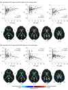
The regression model that tested CPF exposure effects at each white matter voxel was DTI measure = β0 + β1 × CPF + β2 × age + β3 × sex + β4 × race/ethnicity + β5 × maternal education + β6 × material hardship + β7 × home stress + ε, with DTI measure either fractional anisotropy (FA) or average diffusion coefficient (ADC). The statistical significance (cluster size familywise error rate [FWER]–corrected P values) of the associations of exposure with cortical thickness at each point on the surface of the brain are color-coded. Only P values that survived cluster-size FWER correction are plotted. The number of participants in this analysis was 202 (mean [SD] age, 10.78 [1.33] years; 89 boys and 113 girls). The z values below each column represent the z coordinate in Talairach space. FA and ADC values were sampled at representative points, and scatterplots for the association of CPF exposure with the DTI values at those points are shown. DTI values are adjusted for participant age at magnetic resonance imaging scan, sex, race, ethnicity, maternal education, material hardship during pregnancy, and home stress at child age 3 years. IC indicates internal capsule; ILF, inferior longitudinal fasciculus; SLF, superior longitudinal fasciculus.
Figure 4.
Figure 4.. Statistical Maps of Prenatal Chlorpyrifos (CPF) Exposure Associations With Arterial Spin Labeling (ASL) Regional Cerebral Blood Flow (rCBF) Measures
The regression model that tested CPF exposure effects voxelwise was rCBF = β0 + β1 × CPF + β2 ×age + β3 × sex + β4 × race/ethnicity + β5 × maternal education + β6 × material hardship + β7 × home stress + ε. The statistical significance (cluster size familywise error rate [FWER]–corrected P values) of the associations of exposure with cortical thickness at each point on the surface of the brain are color-coded. Only P values that survived cluster-size FWER correction are plotted. The number of participants for ASL was 175 (mean [SD] age, 10.89 [1.24] years; 75 boys and 100 girls). The z values below each column represent the z coordinate in Talairach space. rCBF data were sampled at representative points. Scatterplots showing the association of CPF exposure with those measures are shown for those points. rCBF values are adjusted for participant age at magnetic resonance imaging scan, sex, race, ethnicity, maternal education, material hardship during pregnancy, and home stress at child age 3 years. Because the significant associations for rCBF involved so much of the brain, we show the T1-weighted anatomical template brain for the corresponding slices and the locations where we sampled the ASL data immediately above the statistical maps. aCR indicates anterior corona radiata; IFO, inferior fronto-occipital fasciculus; Ins, insula; PTR, posterior thalamic radiation; SLF, superior longitudinal fasciculus.

References

    1. Solomon KR, Williams WM, Mackay D, Purdy J, Giddings JM, Giesy JP. Properties and uses of chlorpyrifos in the United States. Rev Environ Contam Toxicol. 2014;231:13-34. doi: 10.1007/978-3-319-03865-0_2 - DOI - PubMed
    1. Gibbs JL, Yost MG, Negrete M, Fenske RA. Passive sampling for indoor and outdoor exposures to chlorpyrifos, azinphos-methyl, and oxygen analogs in a rural agricultural community. Environ Health Perspect. 2017;125(3):333-341. doi: 10.1289/EHP425 - DOI - PMC - PubMed
    1. Saunders M, Magnanti BL, Correia Carreira S, et al. Chlorpyrifos and neurodevelopmental effects: a literature review and expert elicitation on research and policy. Environ Health. 2012;11(suppl 1):S5. doi: 10.1186/1476-069X-11-S1-S5 - DOI - PMC - PubMed
    1. Morgan MK, Sheldon LS, Croghan CW, et al. Exposures of preschool children to chlorpyrifos and its degradation product 3,5,6-trichloro-2-pyridinol in their everyday environments. J Expo Anal Environ Epidemiol. 2005;15(4):297-309. doi: 10.1038/sj.jea.7500406 - DOI - PubMed
    1. Bradman A, Barr DB, Claus Henn BG, Drumheller T, Curry C, Eskenazi B. Measurement of pesticides and other toxicants in amniotic fluid as a potential biomarker of prenatal exposure: a validation study. Environ Health Perspect. 2003;111(14):1779-1782. doi: 10.1289/ehp.6259 - DOI - PMC - PubMed