. 2025 Aug 18;20(8):e0328139.
doi: 10.1371/journal.pone.0328139. eCollection 2025.

Curcumin inhibits ferroptosis through dessuccinylation of SIRT5-associated ACSL4 protein, and plays a chondroprotective role in osteoarthritis

Affiliations

Curcumin inhibits ferroptosis through dessuccinylation of SIRT5-associated ACSL4 protein, and plays a chondroprotective role in osteoarthritis

Yong Xu et al. PLoS One. .

Abstract

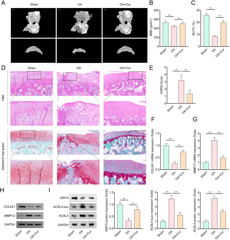
Background: Ferroptosis of chondrocytes plays a crucial role in the progression of osteoarthritis (OA). This study aimed to explore the role of curcumin (Cur) in interfering with chondrocyte ferroptosis in OA.

Methods: Rat chondrocytes were treated with 10 ng/mL interleukin-1β (IL-1β) for 48 hours to mimic the OA microenvironment. The protective effects of Cur were evaluated in vitro by assessing cell viability and ferroptosis. Molecular docking was performed to validate the structural interaction between Cur and the SIRT5 protein. Co-immunoprecipitation (CO-IP) confirmed the binding relationship between SIRT5 and ACSL4. Additionally, the efficacy of Cur in alleviating OA progression was assessed in an in vivo OA rat model.

Results: Cur treatment significantly attenuated IL-1β-induced chondrocyte injury by enhancing cell viability and inhibiting ferroptosis. Cur also markedly reduced global protein lysine succinylation levels. IL-1β suppressed SIRT5 expression, while Cur treatment upregulated SIRT5 expression. The molecular structure of Cur exhibits strong complementarity with the SIRT5 protein, forming a stable complex with high binding affinity. Inhibition of SIRT5 attenuated the protective effects of Cur on chondrocytes and increased ACSL4 succinylation levels. SIRT5 physically interacted with ACSL4, and SIRT5-mediated desuccinylation of ACSL4 repressed its function, thereby mitigating ferroptosis. Cur alleviates OA progression in vivo by inhibiting cartilage destruction, bone erosion, and chondrocyte injury, and by smoothing subchondral bone surfaces.

Conclusion: Cur protects chondrocytes in vitro by inhibiting ferroptosis and suppresses cartilage degeneration and bone erosion in vivo, demonstrating a chondroprotective role in OA. These effects are mediated through SIRT5-dependent desuccinylation of ACSL4, which regulates ferroptosis pathways.


Conflict of interest statement

The authors have declared that no competing interests exist.

Figures

Fig 1
Fig 1. Cur treatment protects chondrocytes from IL-1β induced injury by promoting cell viability and inhibiting cell ferroptosis.
(A) The chemical structural formula of Cur. (B) Cell viability of chondrocytes obtained by CCK-8 method. Cells were treated with 10 μM Cur for 12 hours hours prior to IL-1β stimulation (10 ng/mL, 48 hours) and then incubated with CCK-8 solution. The absorbance at 450 nm was measured using a microplate reader to quantify viable cells, n = 3. (C–D) DAPI/PI staining method was applied to evaluate cell death of condrocytes. After treatment, cells were fixed, permeabilized, and stained with DAPI and PI. Images were captured using fluorescence microscopy, n = 3. (red: PI-labeled dead cells; blue: DAPI-labeled nuclei). (E–H) Cell ferroptosis of chondrocytes was accessed by evaluating ROS, MDA, GSH, and Fe2+ levels. Intracellular ROS levels were measured using DCFH-DA fluorescence. MDA levels were determined through a thiobarbituric acid assay. GSH levels were measured using an enzymatic recycling method. Fe2+ levels were quantified with a ferrozine-based assay, n = 3. (I) Representative protein bands and quantitative analysis of GPX4 and ACSL4 levels in chondrocytes. Protein samples were extracted, separated by SDS-PAGE, transferred to PVDF membranes, and incubated with specific antibodies against GPX4 and ACSL4. Band intensities were analyzed using ImageJ software, n = 3. Data were presented as mean ± SD. Comparisons between groups were performed using one-way ANOVA followed by Tukey’s post-hoc test. **p < 0.01.
Fig 2
Fig 2. Cur reduces global protein succinylation and upregulates SIRT5 expression, with molecular docking suggesting a direct interaction between Cur and SIRT5.
(A) The representative protein images and quantitative analysis of total succinylation of lysine of proteins in chondrocytes. Cells were lysed, protein samples were prepared and subjected to Western blot analysis using anti-succinyllysine antibodies, n = 3. (B) The representative protein images and quantitative analysis of protein levels of four succinylase (KAT2A, KAT3B, CPT1A, and HAT1) and two desuccinylase (SIRT5 and SIRT7) in chondrocytes of each group. Protein levels were determined using Western blot assay with specific antibodies, n = 3. (C) The enzyme activity of SIRT5 in chondrocytes before and after Cur treatment. SIRT5 activity was measured using a commercially available SIRT5 activity assay kit according to the manufacturer’s instructions, n = 3. (D) The molecular docking results suggested that structure of Cur compound is well matched with SIRT5 target protein. Docking studies were performed using molecular docking software to predict the binding mode and affinity between Cur and SIRT5. Data were presented as mean ± SD. Comparisons between groups were performed using one-way ANOVA followed by Tukey’s post-hoc test. **p < 0.01.
Fig 3
Fig 3. Inhibition of SIRT5 weakens the protecting effects of Cur on chondrocytes.
(A) PCR and western blotting analysis were performed to evaluate the levels of SIRT5 in chondrocytes transfected with shNC or shSIRT5. Total RNA was extracted, reverse-transcribed into cDNA, and amplified using specific primers for SIRT5. Protein samples were also collected and subjected to Western blot analysis with SIRT5-specific antibodies, n = 3. (B) Cell viability of chondrocytes transfected with shNC or shSIRT5 were obtained by CCK-8 method after IL-1β treatment, n = 3. (C) DAPI/PI staining method was applied to evaluate cell death of IL-1β treated chondrocytes (transfected with shNC or shSIRT5), n = 3. (red: PI-labeled dead cells; blue: DAPI-labeled nuclei). (D–G) Cell ferroptosis of IL-1β treated chondrocytes (transfected with shNC or shSIRT5)was accessed by evaluating ROS, MDA, GSH, and Fe2+ levels, n = 3. (H) Representative protein bands and quantitative analysis of GPX4 and ACSL4 levels in IL-1β treated chondrocytes (transfected with shNC or shSIRT5), n = 3. Data were presented as mean ± SD. Comparisons between groups were performed using Student’s t-test or one-way ANOVA followed by Tukey’s post-hoc test. *p < 0.05, **p < 0.01.
Fig 4
Fig 4. SIRT5 interacts with ACSL4 and regulates its succinylation status.
(A) Western blot was carried out to evaluate the succinylation level of ferroptosis-related proteins in chondrocytes transfected with shNC or shSIRT5, n = 3. (B) CO-IP was performed to verify the endogenous binding relationship between SIRT5 and ACSL4. Cell lysates were incubated with SIRT5-specific antibodies and protein A/G agarose beads. The immunoprecipitated proteins were analyzed by Western blot using ACSL4-specific antibodies, n = 3. (C) The succinylation level of ACSL4 protein in chondrocytes (transfected with shNC or shSIRT5) after muation at the predicted succinylation sites, n = 3. (D) Quantitative analysis of protein degradation of ACSL4 when SIRT5 was inhibited. Protein levels of ACSL4 were determined at different time points after inhibiting SIRT5 expression, n = 3. Data were presented as mean ± SD. Comparisons between groups were performed using Student’s t-test. *p < 0.05, **p < 0.01.
Fig 5
Fig 5. SIRT5-mediated cell ferroptosis is reversed by the elevation of ACSL4.
(A–B) PCR and western blotting analysis were performed to evaluate the levels of SIRT5 and ACSL4 in chondrocytes transfected with vector or SIRT5. Total RNA and protein samples were collected, and SIRT5 and ACSL4 levels were determined using PCR and Western blotting, n = 3. (C) Cell viability of IL-1β treated chondrocytes (transfected with vector or SIRT5) obtained by CCK-8 method, n = 3. (D) DAPI/PI staining method was applied to evaluate cell death of IL-1β treated chondrocytes (transfected with vector or SIRT5), n = 3. (red: PI-labeled dead cells; blue: DAPI-labeled nuclei). (E–H) Cell ferroptosis of IL-1β treated chondrocytes (transfected with vector or SIRT5) was accessed by evaluating ROS, MDA, GSH, and Fe2+ levels, n = 3. (I) Representative protein bands and quantitative analysis of GPX4 and ACSL4 levels in IL-1β treated chondrocytes (transfected with vector or SIRT5), n = 3. Data were presented as mean ± SD. Comparisons between groups were performed using Student’s t-test or one-way ANOVA followed by Tukey’s post-hoc test. *p < 0.05, **p < 0.01.
Fig 6
Fig 6. Cur alleviate OA progress in vivo.
(A) Representative CT images of the femurs of rats. Micro-CT scanning was performed to visualize the bone structure of rat femurs. (B–C) Quantitative analysis of bone mineral density (BMD), and bone volume to tissue volume (BV/TV) were measured. BMD and BV/TV values were calculated from Micro-CT images, n = 6. (D) Representative images of H&E staining and Saffron O-solid green staining (red indicates cartilage tissues, and green indicates mineralized bone tissue sites). (E) The OARSI score assessed the degree of articular cartilage injury and the progression of OA. OARSI scoring was performed by blinded observers using a standardized scoring system, n = 6. (F–G) PCR analysis was performed to evaluate the mRNA levels of COL2A1 and MMP13 in cartilage tissues, n = 6. (H) Western blot analysis was performed to evaluate the protein levels of COL2A1 and MMP13 in cartilage tissues, n = 6. (I) Representative protein bands and quantitative analysis of SIRT5 and ACSL4 levels in cartilage tissues, n = 6. Data were presented as mean ± SD. Comparisons between groups were performed using one-way ANOVA followed by Tukey’s post-hoc test. **p < 0.01.

Similar articles

References

    1. Motta F, Barone E, Sica A, Selmi C. Inflammaging and osteoarthritis. Clin Rev Allergy Immunol. 2023;64(2):222–38. doi: 10.1007/s12016-022-08941-1 - DOI - PubMed
    1. Kim HL, Lee HJ, Lee D, Choi B, Yang SH. Herbal composition LI73014F2 alleviates articular cartilage damage and inflammatory response in monosodium iodoacetate-induced osteoarthritis in rats. Molecules. 2020;25(22):5467. doi: 10.3390/molecules25225467 - DOI - PMC - PubMed
    1. Zhou X, Zheng Y, Sun W, Zhang Z, Liu J, Yang W. D-mannose alleviates osteoarthritis progression by inhibiting chondrocyte ferroptosis in a HIF-2alpha-dependent manner. Cell Prolif. 2021;54(11):e13134. doi: 10.1111/cpr.13134 - DOI - PMC - PubMed
    1. Sun K, Hou L, Guo Z, Wang G, Guo J, Xu J, et al. JNK-JUN-NCOA4 axis contributes to chondrocyte ferroptosis and aggravates osteoarthritis via ferritinophagy. Free Radic Biol Med. 2023;200:87–101. doi: 10.1016/j.freeradbiomed.2023.03.008 - DOI - PubMed
    1. Chen Y, Liu Y, Jiang K, Wen Z, Cao X, Wu S. Linear ubiquitination of LKB1 activates AMPK pathway to inhibit NLRP3 inflammasome response and reduce chondrocyte pyroptosis in osteoarthritis. J Orthop Translat. 2023;39:1–11. doi: 10.1016/j.jot.2022.11.002 - DOI - PMC - PubMed

LinkOut - more resources