

Marine Autographiviridae phages exhibit high genetic diversity and global-scale biogeographic patterns
- PMID: 40825829
- PMCID: PMC12361551
- DOI: 10.1038/s42003-025-08611-w
Marine Autographiviridae phages exhibit high genetic diversity and global-scale biogeographic patterns
Abstract
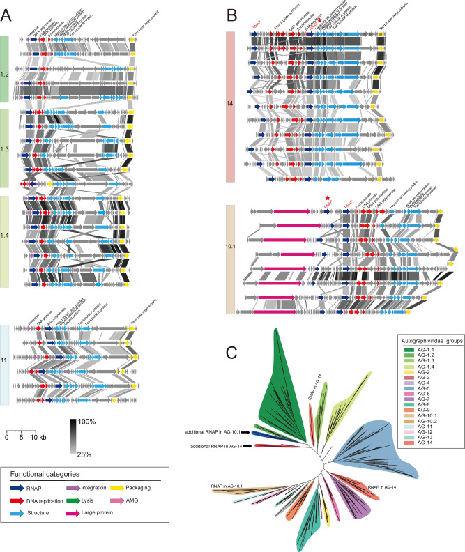
Marine viral communities harbor an astounding diversity of phages infecting diverse marine bacteria. The double-stranded DNA phage family Autographiviridae is among the most abundant phage families in the ocean. However, the current understanding of marine Autographiviridae phages is predominantly derived from isolates infecting cyanobacteria, SAR11, and Roseobacter. To achieve a more comprehensive understanding of the diversity, ecological traits, and functional profiles of this phage family, we recovered 1253 complete marine Autographiviridae uncultivated viral genomes (UViGs). Comparative genomic analysis showed that marine-derived Autographiviridae phages display genome synteny and share a conserved core of seven genes. The core gene-based phylogenomic analysis classified them into 14 groups, 6 of which were previously undescribed. These groups varied in G + C content, genome size, and presence of specific genes. Metagenomic recruitment analysis demonstrated that Autographiviridae phages are globally distributed and enriched in the upper ocean layers of tropical and temperate zones. The differential distribution patterns among these groups mirror the ecological niches of their potential hosts, emphasizing the top-down control these phages exert on their host populations. Collectively, our study substantially expands knowledge regarding the diversity, potential hosts, functional capacity, and ecological distribution of Autographiviridae phages in the ocean, emphasizing their ecological implications in marine environments.
© 2025. The Author(s).
Conflict of interest statement
Competing interests: The authors declare no competing interests.
Figures


Similar articles
-
Horizontal Gene Transfer and CRISPR Targeting Drive Phage-Bacterial Host Interactions and Coevolution in "Pink Berry" Marine Microbial Aggregates.Appl Environ Microbiol. 2023 Jul 26;89(7):e0017723. doi: 10.1128/aem.00177-23. Epub 2023 Jul 5. Appl Environ Microbiol. 2023. PMID: 37404190 Free PMC article.
-
Genome sequences of the first Autographiviridae phages infecting marine Roseobacter.Microb Genom. 2024 Apr;10(4):001240. doi: 10.1099/mgen.0.001240. Microb Genom. 2024. PMID: 38630615 Free PMC article.
-
Novel phage infecting the Roseobacter CHUG lineage reveals a diverse and globally distributed phage family.mSphere. 2024 Jul 30;9(7):e0045824. doi: 10.1128/msphere.00458-24. Epub 2024 Jun 27. mSphere. 2024. PMID: 38926906 Free PMC article.
-
Marine Bacteriophages as Next-Generation Therapeutics: Insights into Antimicrobial Potential and Application.Viruses. 2025 Jul 10;17(7):971. doi: 10.3390/v17070971. Viruses. 2025. PMID: 40733588 Free PMC article. Review.
-
The quantity, quality and findings of network meta-analyses evaluating the effectiveness of GLP-1 RAs for weight loss: a scoping review.Health Technol Assess. 2025 Jun 25:1-73. doi: 10.3310/SKHT8119. Online ahead of print. Health Technol Assess. 2025. PMID: 40580049 Free PMC article.
References
-
- Fuhrman, J. A. Marine viruses and their biogeochemical and ecological effects. Nature399, 541–548 (1999). - PubMed
-
- Suttle, C. A. Viruses in the sea. Nature437, 356–361 (2005). - PubMed
-
- Suttle, C. A. Marine viruses-major players in the global ecosystem. Nat. Rev. Microbiol.5, 801–812 (2007). - PubMed
-
- Breitbart, M. Marine viruses: truth or dare. Annu. Rev. Mar. Sci.4, 425–448 (2012). - PubMed
MeSH terms
Grants and funding
LinkOut - more resources
Full Text Sources
