Review
. 2025 Aug 18;29(1):362.
doi: 10.1186/s13054-025-05586-2.

Neuroimaging for neurovascular complications of traumatic brain injury

Affiliations
Review

Neuroimaging for neurovascular complications of traumatic brain injury

Laura Saitta et al. Crit Care. .

Abstract

Background: Traumatic brain injury typically causes extra-axial and/or intra-axial bleeding including subarachnoid hemorrhage, intraparenchymal hemorrhage, subdural hematomas and epidural hematomas. Less commonly, trauma can cause cerebrovascular complications, which involve either the arterial or the venous side. Because of the rarity of these pathological conditions, guidelines and recommendations for their management are still controversial.

Main body: The objective of this work is to describe the possible cerebrovascular complications of critically ill traumatic brain injured patients and to understand the most common underlying mechanisms and radiological features as well as their management. A variety of pathological entities will be addressed, such as post-traumatic aneurysms, carotid-cavernous fistula, arterial occlusion, arterial dissection (in potential association with brain ischemia), as well as arterial rupture/avulsion and post-traumatic venous thrombosis. Neurovascular complications of head trauma vary depending on the traumatic mechanism, on the site of impact and on the osseous structures involved. Early diagnosis is mostly based on Computed Tomography/Computed Tomography Angiography (CT/CTA) whose findings help guide patient management by detecting vascular lesions potentially leading to neurological deterioration. Magnetic resonance imaging may be useful in selected cases. Today Digital Subtraction Angiography (DSA) is mostly a diagnostic problem-solving tool when CTA findings are equivocal but advanced endovascular interventional techniques have improved the therapeutic possibilities in post-traumatic vascular complications. CONCLUSIONS: Neurovascular complications are not common after head trauma but should not be overlooked because they might lead to severe and life-threatening consequences. Early diagnosis, and a multidisciplinary collaboration including neuroradiologists, neurosurgeons and neurointensivists is fundamental in order to prevent and minimize secondary brain damage in this population.

Keywords: Neurocritical care; Neuroimaging; Neurovascular complications; Traumatic brain injury.


Conflict of interest statement

Declarations. Ethics approval and consent to participate: The ethics committee approved the study (reference number register CER Liguria: 340/2022—DB id 12,413). Consent for publication: Not applicable. Competing interests: The authors declare no competing interests.

Figures

Fig. 1
Fig. 1
Anatomy of the osseous structures and foramina observed in the anterior, middle and posterior cranial fossa, with respect to the vascular structures which could possibly be injured when involved in TBI
Fig. 2
Fig. 2
Schematic drawing of the five grades of vascular injury according to the Denver scale. Grade I may present as irregularity of the vessel wall [1], subtle intimal flap [2] or intramural hematoma with < 25% stenosis; grade II may present as frank intimal flap [1], intramural hematoma with > 25% stenosis [2] or intraluminal thrombus [3]; grade III is represented by the presence of a pseudoaneurysm; grade IV occurs in case of arterial occlusion by a complete intimal flap [1] or by an intramural hematoma [2]; grade V is represented by arterial transection
Fig. 3
Fig. 3
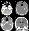
Post-traumatic bilateral internal carotid dissection and left carotid fusiform dilatation in an adolescent patient following a motor vehicle accident. CT scan shows a parieto-temporal epidural hemorrhage (A) in association with a skull fracture at the same level (B, arrow). CTA demonstrates a bilateral double lumen sign (C, arrows), consistent with the presence of intimal flap and a distinct narrowing of the left vertical petrous carotid (D, arrowhead). Maximum intensity projection (E) and 3D volume rendering (F, arrowheads) show the presence of a dilatation of the left carotid artery (consistent with a pseudoaneurysm due to a tear of the intima and hemorrhage into media and adventitia layers)
Fig. 4
Fig. 4
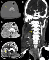
Post-traumatic cervical CT, CTA and MRI of a patient in the sixth decade of life involved in a car accident. Non-enhanced CT scan shows bilateral fractures of the lateral masses of a cervical vertebra (A), with involvement of the transverse foramina. CTA (B) shows partial lack of opacification of the left vertebral artery, consistent with post-traumatic arterial dissection (arrow) (Denver grade II). The MRI (C) shows foci of restricted diffusion in the diffusion weighted sequence in the right cerebellum, consistent with acute ischemic lesions of embolic origin from the left vertebral artery
Fig. 5
Fig. 5
Basal CT scan of a patient in the early fifth decade of life following a motor vehicle accident. Non-enhanced CT scan (A) demonstrates a fracture of the right superior articular process of the C6 vertebra (yellow arrowhead). CTA (B, C) shows lack of opacification of the right vertebral artery in the transverse foramen (B, yellow arrowhead), starting from the distal V1 segment (not shown), continuing along part of the V2 segment (C, black arrowheads) and V3 segment and ending at the proximal V4 segment (not shown), consistent with arterial dissection with subsequent occlusion of the vessel (Denver Grade IV)
Fig. 6
Fig. 6
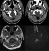
Blunt trauma in a patient at the beginning of the third decade of life involved in a motor vehicle accident. MRI (top row) shows areas of restricted diffusion (A: diffusion weighted imaging sequence and B: apparent diffusion coefficient map) in the centrum semiovale of the right cerebral hemisphere, consistent with acute ischemia. MRA (C) demonstrates distinct narrowing of the right intracranial carotid artery (arrow) involving the cavernous and supraclinoid segments, consistent with post-traumatic dissection. Follow-up CTA one month from presentation (D-F) shows the appearance of a bilobated post-traumatic aneurysm (Denver grade III) of the right internal carotid artery, with a component arising from the supraclinoid tract and directed supero-laterally (D, arrow) and a component arising from the cavernous portion and directed medially in the sellar region (E, arrowhead). Maximum intensity projection reconstruction (F) more clearly depicts the supraclinoid portion of the aneurysm (arrow)
Fig. 7
Fig. 7
Right vertebral artery rupture after motor vehicle collision in a patient in the seventh decade of life. Non-enhanced CT scan (A and B) at the level of C5 shows fracture and lateral dislocation of the right transverse process (arrow), with ipsilateral distinct collection in the soft tissues of the neck (B). CTA (C) shows contrast extravasation in the right lateral cervical soft tissues, consistent with acute hematoma (asterisk), with lack of opacification of the right vertebral artery, best seen in the maximum intensity projection reconstruction (D, dotted arrows), most consistent with vertebral artery avulsion
Fig. 8
Fig. 8
CTA and MRI findings of arterial dissection in the presence of a mural hematoma. CTA (top row) shows a slight lumen reduction of the distal cervical segment of the left internal carotid artery, where an intramural hematoma is also suspected, showing isodensity with the surrounding soft tissues (arrowheads). MRI (bottom row) at the same level more clearly depicts a “crescent sign” consistent with a subacute intramural hematoma on T1-weighted (bottom left) and on DWI (bottom right) sequences (arrowheads)
Fig. 9
Fig. 9
Patient in the sixth decade of life with a penetrating spear gun harpoon trauma (A, scout image). The harpoon penetrated the brain through the hard palate and the cranial base, with absence of significant cerebral hemorrhagic hyperdensity on non-enhanced CT (B), and a possible slight narrowing of the left distal middle cerebral artery on CTA (C), in the presence of metal artifact. Bottom row shows the post-surgical control, after removal of the harpoon, demonstrating the occurrence of ventricular hemorrhage and left intraparenchymal hematoma on non-enhanced CT (D), with evidence of a pseudoaneurysm arising from the supraclinoid left internal carotid artery on CTA (E and F, arrows)
Fig. 10
Fig. 10
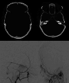
Blunt trauma in a patient in the early eighth decade of life. CT scan (top row) shows a non-otic longitudinal fracture of the right petrous bone. DSA (bottom row) performed while injecting the right external carotid artery territories, demonstrates the presence of a post-traumatic dural AVF between the posterior branch of the middle meningeal artery and the ipsilateral transverse sinus (Borden type I)
Fig. 11
Fig. 11
Post-traumatic direct CCF of a patient in the late sixth decade of life following a car accident. Non-enhanced CT scan (A, B) shows the presence of minimal subarachnoid hemorrhage in interpeduncular cistern and a small temporo-mesial intraparenchymal hemorrhage (A), with multiple fractures involving the left zygoma, the left maxillary sinus (B) and the left mandibular angle (not shown). CTA (C) shows prominent opacification and bulging of the cavernous sinus on both sides (asterisks) and bilateral enlargement of the superior ophtalmic veins (arrows). DSA (middle and bottom row) was performed two days later and showed a left CCF (D, E, F: lateral view; G, H: frontal view) with evidence of venous drainage via the anterior route, via the postero-inferior route and with cortical drainage through the left spheno-parietal sinus. Occlusion of the CCF was obtained with a detachable balloon (I, yellow arrow)
Fig. 12
Fig. 12
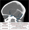
Schematic representation of the types of venous drainage
Fig. 13
Fig. 13
VAVF of an adolescent patient with neurofibromatosis 1 after a minor trauma. In A (coronal reconstructions) and B (axial native images) CTA shows asymmetric opacification of the cervical venous plexus, with early enhancement in the left side. In C, D, E and F the pre-operative DSA, performed while injecting the left vertebral artery, better demonstrates the presence of a VAVF between the left vertebral artery and the left cervical venous plexus, with evidence of dilated and tortuous arterialized veins (C, E: frontal views. D, F: lateral views). In G (frontal view) and H (lateral view) post-treatment results are shown, with complete occlusion of the fistula by coiling and preservation of the parent artery patency in the arterial phase
Fig. 14
Fig. 14
Blunt trauma in a patient at the beginning of the ninth decade of life. Non-enhanced CT scan (A, B) obtained at presentation shows the presence of air bubbles and hyperdensity of the right transverse sinus (A, circle), adjacent to a diastasis of the occipito-mastoid suture, with a fracture of the ipsilateral squamous occipital bone (B, arrows). Follow-up CTA performed after 48 h (C, D), shows a lack of opacification of the right transverse sinus (C, dotted arrow) and of the ipsilateral jugular vein (D, arrowhead), consistent with venous thrombosis
Fig. 15
Fig. 15
Blunt trauma in a patient in their twenties. Non-enhanced brain CT shows a post-traumatic fracture of the frontal bone (A), with evidence of an extra-axial blood collection, consistent with midline epidural hematoma of the cranial vault (B). CTV (C) demonstrates lack of opacification of the superior sagittal sinus, resulting from compression by the epidural hematoma
Fig. 16
Fig. 16
CT scan of a young adult patient involved in a motor vehicle accident. A post-traumatic diastasis of the left occipito-mastoid suture is shown (A, arrowhead) with hyperdensity of the left sigmoid sinus (B, arrowhead). A fracture of the left anterior occipital bone, posterior to the ipsilateral hypoglossal canal is also shown (A). The CTV (C) shows lack of opacification of the left sigmoid sinus, consistent with venous thrombosis (arrowhead)
Fig. 17
Fig. 17
CT scan of a patient in the early sixth decade of life with a depressed skull fracture of the parietal bone (A), with findings consistent with thrombosis of a cortical vein. Non-contrast CT scan (B) shows a linear hyperdensity at the level of the right post-central sulcus (yellow arrows) along the presumed anatomical course of a cortical vein, adjacent to the fracture. Follow-up CTV at 48 h (C), shows absent/irregular opacification along the course of a right parietal cortical vein (arrows), corresponding to the non-contrast finding, consistent with cortical vein thrombosis. The superior sagittal sinus (not shown) was patent

Similar articles

  • Prescription of Controlled Substances: Benefits and Risks.
    Preuss CV, Kalava A, King KC. Preuss CV, et al. 2025 Jul 6. In: StatPearls [Internet]. Treasure Island (FL): StatPearls Publishing; 2025 Jan–. 2025 Jul 6. In: StatPearls [Internet]. Treasure Island (FL): StatPearls Publishing; 2025 Jan–. PMID: 30726003 Free Books & Documents.
  • Elbow Fractures Overview.
    Waseem M, Saeed W, Launico MV. Waseem M, et al. 2025 Jul 7. In: StatPearls [Internet]. Treasure Island (FL): StatPearls Publishing; 2025 Jan–. 2025 Jul 7. In: StatPearls [Internet]. Treasure Island (FL): StatPearls Publishing; 2025 Jan–. PMID: 28723005 Free Books & Documents.
  • Sexual Harassment and Prevention Training.
    Cedeno R, Bohlen J. Cedeno R, et al. 2024 Mar 29. In: StatPearls [Internet]. Treasure Island (FL): StatPearls Publishing; 2025 Jan–. 2024 Mar 29. In: StatPearls [Internet]. Treasure Island (FL): StatPearls Publishing; 2025 Jan–. PMID: 36508513 Free Books & Documents.
  • Management of urinary stones by experts in stone disease (ESD 2025).
    Papatsoris A, Geavlete B, Radavoi GD, Alameedee M, Almusafer M, Ather MH, Budia A, Cumpanas AA, Kiremi MC, Dellis A, Elhowairis M, Galán-Llopis JA, Geavlete P, Guimerà Garcia J, Isern B, Jinga V, Lopez JM, Mainez JA, Mitsogiannis I, Mora Christian J, Moussa M, Multescu R, Oguz Acar Y, Petkova K, Piñero A, Popov E, Ramos Cebrian M, Rascu S, Siener R, Sountoulides P, Stamatelou K, Syed J, Trinchieri A. Papatsoris A, et al. Arch Ital Urol Androl. 2025 Jun 30;97(2):14085. doi: 10.4081/aiua.2025.14085. Epub 2025 Jun 30. Arch Ital Urol Androl. 2025. PMID: 40583613 Review.
  • The Black Book of Psychotropic Dosing and Monitoring.
    DeBattista C, Schatzberg AF. DeBattista C, et al. Psychopharmacol Bull. 2024 Jul 8;54(3):8-59. Psychopharmacol Bull. 2024. PMID: 38993656 Free PMC article. Review.

References

    1. Weber CD, Lefering R, Kobbe P, Horst K, Pishnamaz M, Sellei RM, et al. Blunt cerebrovascular artery injury and stroke in severely injured patients: an international multicenter analysis. World J Surg. 2018;42(7):2043–53. - PubMed
    1. Esnault P, Cardinale M, Boret H, D’Aranda E, Montcriol A, Bordes J, et al. Blunt cerebrovascular injuries in severe traumatic brain injury: incidence, risk factors, and evolution. J Neurosurg. 2017;127(1):16–22. - PubMed
    1. Krings T, Geibprasert S, Lasjaunias PL. Cerebrovascular trauma. Eur Radiol. 2008;18(8):1531–45. - PubMed
    1. Stone DK, Viswanathan VT, Wilson CA. Management of blunt cerebrovascular injury. Curr Neurol Neurosci Rep. 2018;18(12):1–9. - PubMed
    1. Steenburg SD, Sliker CW, Shanmuganathan K, Siegel EL (2010) Imaging evaluation of penetrating neck injuries. Radiographics 30:869–886 - PubMed

LinkOut - more resources