. 2025 Aug 13:18:10969-10994.
doi: 10.2147/JIR.S532630. eCollection 2025.

Mechanism of Lipi Jiangzhuo Decoction in Improving Metabolic Dysfunction-Associated Steatohepatitis Through the PERK/PINK1/GPx4 Pathway

Affiliations

Mechanism of Lipi Jiangzhuo Decoction in Improving Metabolic Dysfunction-Associated Steatohepatitis Through the PERK/PINK1/GPx4 Pathway

Linlin Pan et al. J Inflamm Res. .

Abstract

Background: Metabolic dysfunction-associated steatohepatitis (MASH) is characterized primarily by hepatocyte lipoapoptosis and hepatic inflammation, frequently developing from overweight/obesity. To date, no specific therapeutics exist to reverse MASH. Although resmetirom has been approved in some regions, patients in many Asian countries, including China, still lack access to approved pharmacotherapy for MASH. Lipi Jiangzhuo decoction (LPJZD) is a promising traditional Chinese medicine formula for MASH. However, to date, there have been no comprehensive studies clarifying its potential mechanism of action. This study aims to elucidate the underlying mechanism of action of LPJZD in the treatment of MASH.

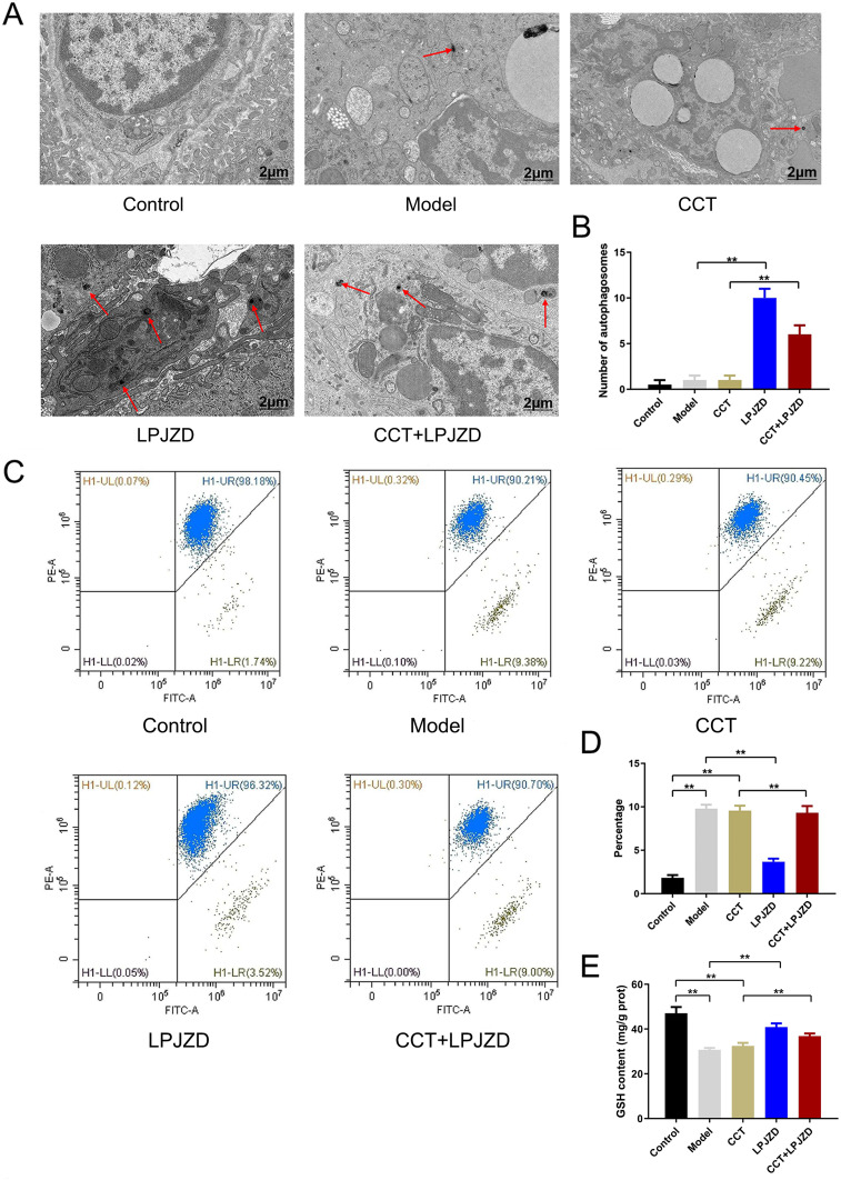
Materials and methods: A MASH mouse model was established by feeding a high-fat diet and subjecting them to fatigue protocols and cold stress for 12 weeks. After treating MASH mice with LPJZD, biochemical assays were conducted to assess the efficacy of LPJZD in alleviating the MASH symptoms. In addition, the in vitro effects of LPJZD on MASH were evaluated using L-02 cells. Specifically, we analyzed the effect of LPJZD on endoplasmic reticulum (ER) stress, mitophagy, and ferroptosis by Western blot analysis, flow cytometry, immunofluorescence staining, and enzyme-linked immunosorbent assay.

Results: In vivo, LPJZD effectively improved the inflammatory response, reduced body weight and blood lipid levels, improved liver function, reduced liver lipid droplet accumulation, and ameliorated the pathological status of MASH mice. In vitro, LPJZD effectively inhibited ferroptosis by reducing ferrous ions and reactive oxygen species levels, increasing GPx4 protein expression, elevating glutathione levels, and ameliorating mitochondrial swelling and matrix thinning. Simultaneously, LPJZD activated mitophagy by increasing PINK1 and Parkin protein expression, augmenting mitophagosome number, and restoring mitochondrial membrane potential. Additionally, LPJZD suppressed ER stress by decreasing PERK protein expression. Notably, activation of ER stress using a PERK activator attenuated LPJZD's effects on mitophagy activation and ferroptosis inhibition, inhibition of mitophagy via a PINK1 inhibitor diminished LPJZD's anti-ferroptotic effect, and administration of a GPx4 inhibitor reduced LPJZD's suppression of ferroptosis. Therefore, these results demonstrate that LPJZD ameliorates MASH by regulating the PERK/PINK1/GPx4 pathway.

Conclusion: LPJZD can improve MASH by regulating ER stress-mitophagy -ferroptosis axis in liver cells. The role of LPJZD in anti-inflammatory therapy provides new insights for the clinical prevention and treatment of MASH.

Keywords: Lipi Jiangzhuo decoction; endoplasmic reticulum stress; ferroptosis; metabolic dysfunction-associated steatohepatitis; mitophagy.


Conflict of interest statement

The authors declare that the research was conducted in the absence of any commercial or financial relationships that could be construed as a potential conflict of interest.

Figures

None
Graphical abstract
Figure 1
Figure 1
Identification of chemical compounds in the LPJZD.
Figure 2
Figure 2
Effects of LPJZD on inflammatory response in the mice with MASH. (A) Changes in body weight during the experiment. (B) The liver weight after treatment. (C) The ratio of liver weight to body weight after treatment. (D) The levels of serum ALT after treatment. (E) The levels of serum AST after treatment. (F) The levels of serum TC after treatment. (G) The levels of serum TG after treatment. (H) The levels of serum HDL-c after treatment. (I) The levels of serum LDL-c after treatment. (J) The levels of MDA after treatment. (K) The levels of SOD after treatment. (L) The levels of IL-6 after treatment. (M) The levels of TNF-α after treatment. (N) Oil-red-O staining was used to visualize lipid droplets in hepatocytes (×400). (O) H&E staining was used to visualize the pathological condition of the liver (×400). A-M: Data from ten independent replicates are presented as the mean ± SD. ** P < 0.01. (NO): Data from three independent replicates are presented as the mean ± SD. ** P < 0.01.
Figure 3
Figure 3
Effects of LPJZD on ER stress, mitophagy, and ferroptosis in the MASH mice. (A) Western blot analysis was performed to determine the protein expression levels of PERK, PINK1, Parkin, and GPx4. (BE): Quantification of protein expression levels of PERK, PINK1, Parkin, and GPx4. Western blot quantification for each indicator was based on sampled from three mice, and the three mice were randomly selected from a corresponding experimental group. Data from three mice are presented as the mean ± SD. ** P < 0.01.
Figure 4
Figure 4
LPJZD inhibits ferroptosis in MASH liver cells. (A) The levels of Fe2+ were detected by immunofluorescence staining (×50). (B) Quantification of the Fe2+ levels. (C) The levels of GPx4 were detected by immunofluorescence staining (×100). (D) Quantification of the GPx4 levels. (E) The levels of ROS were detected by a flow cytometry method using the DCFH-DA fluorescent probe. (F) Quantification of ROS levels. (G) The levels of GSH. Data from three independent replicates are presented as the mean ± SD. ** P < 0.01.
Figure 5
Figure 5
LPJZD inhibits ferroptosis, inflammatory response and apoptosis in MASH liver cells. (A) The morphological changes of mitochondria were observed by scanning electron microscopy (×20000). (B) Evaluation of the effect of different doses of LPJZD on cell viability using MTT assay. (C and D): Evaluation of the effect of different doses of LPJZD on the expression of IL-6 and TNF-α. (E) Evaluation of the effects of LPJZD on apoptosis using flow cytometry. (F) Quantification of apoptosis levels. Data from three independent replicates are presented as the mean ± SD. ** P < 0.01.
Figure 6
Figure 6
LPJZD induces mitophagy in MASH liver cells. (A) Western blot analysis was performed to determine the protein expression levels of PINK1, Parkin, and GPx4. (BD): Quantification of protein expression levels of PINK1, Parkin, and GPx4. (E) Evaluation of the effect of LPJZD on mitochondrial autophagosomes using transmission electron microscopy (×10000). (F) Quantification of the mitophagosome number. (G) Evaluation of the effect of LPJZD on MMP. (H) Quantification of the effect of LPJZD on MMP. Data from three independent replicates are presented as the mean ± SD. ** P < 0.01.
Figure 7
Figure 7
The effect of LPJZD on ferroptosis after inhibition of mitophagy in MASH liver cells. (A) The levels of Fe2+ was determined by immunofluorescence staining (×50). (B) Quantification of the Fe2+ levels. (C) The levels of ROS were determined by flow cytometry using the DCFH-DA fluorescent probe. (D) Quantification of the ROS levels. (E) The levels of GSH. (F) Evaluation of the effect of LPJZD on apoptosis by flow cytometry. (G) Quantification of apoptosis levels. Data from three independent replicates are presented as the mean ± SD. ** P < 0.01.
Figure 8
Figure 8
LPJZD inhibits ER stress in MASH liver cells. (A) Western blot analysis was performed to determine the protein expression levels of PERK, PINK1, Parkin, and GPx4. (BE): Quantification of protein expression levels of PERK, PINK1, Parkin, and GPx4. (F) Evaluation of the effect of LPJZD on GRP78 protein expression using immunofluorescence staining (×100). (G) Quantification of GRP78 protein levels. Data from three independent replicates are presented as the mean ± SD. ** P < 0.01.
Figure 9
Figure 9
Continued.
Figure 9
Figure 9
Effects of LPJZD on mitophagy and ferroptosis in the presence of activated PERK in MASH liver cells. (A) Evaluation of the effect of LPJZD on mitochondrial autophagosomes by transmission electron microscopy (×10000). (B) Quantification of the mitophagosome number. (C) Evaluation of the effect of LPJZD on MMP. (D) Quantification of the effect of LPJZD on MMP. (E) Evaluation of the effect of LPJZD on GSH level. (F) Evaluation of the effect of LPJZD on Fe2+ content by immunofluorescence staining (×50). (G) Quantification of Fe2+ levels. Data from three independent replicates are presented as the mean ± SD. ** P < 0.01.
Figure 10
Figure 10
Schematic representation of the mechanism of action of LPJZD in alleviating MASH.

Similar articles

References

    1. Sanyal AJ, Van Natta ML, Clark J, et al. Prospective study of outcomes in adults with nonalcoholic fatty liver disease. N Engl J Med. 2021;385(17):1559–1569. doi: 10.1056/NEJMoa2029349 - DOI - PMC - PubMed
    1. Powell EE, Wong VW, Rinella M. Non-alcoholic fatty liver disease. Lancet. 2021;397(10290):2212–2224. doi: 10.1016/S0140-6736(20)32511-3 - DOI - PubMed
    1. Zhou F, Zhou J, Wang W, et al. Unexpected rapid increase in the burden of NAFLD in China from 2008 to 2018: a systematic review and meta-analysis. Hepatology. 2019;70(4):1119–1133. doi: 10.1002/hep.30702 - DOI - PubMed
    1. Francque S, Szabo G, Abdelmalek MF, et al. Nonalcoholic steatohepatitis: the role of peroxisomeproliferator-activated receptors. Nat Rev Gastroenterol Hepatol. 2021;18(1):24–39. doi: 10.1038/s41575-020-00366-5 - DOI - PubMed
    1. Younossi ZM, Golabi P, Price JK, et al. The global epidemiology of nonalcoholic fatty liver disease and nonalcoholic steatohepatitis among patients with type 2 diabetes. Clin Gastroenterol Hepatol. 2024;22(10):1999–2010.e8. doi: 10.1016/j.cgh.2024.03.006 - DOI - PubMed

LinkOut - more resources