A cervical spine metastasis of a hepatocellular carcinoma: a rare presentation
- PMID: 40828566
- PMCID: PMC12367086
- DOI: 10.1080/20565623.2025.2548742
A cervical spine metastasis of a hepatocellular carcinoma: a rare presentation
Abstract
Background: Hepatocellular carcinoma (HCC) is a leading cause of cancer-related mortality globally, with bone metastases signifying advanced disease and a poor prognosis. Although bone metastases in HCC are relatively uncommon, they present significant diagnostic and therapeutic challenges. We present a rare case of cervical spine metastasis as a presenting feature of HCC progression, highlighting the need for timely diagnosis and multidisciplinary care.
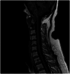
Case presentation: A 55-year-old North African female with a history of hepatitis C-related cirrhosis, successfully treated with antiviral therapy, was diagnosed with HCC following the detection of hepatic nodules during routine biannual surveillance. After unsuccessful radiofrequency ablation (RFA), she developed intense cervical pain. Imaging revealed osteolytic cervical lesions and a paravertebral mass. Biopsy confirmed metastatic HCC. She received radiotherapy and immunotherapy (atezolizumab-bevacizumab). Despite treatment, her condition deteriorated due to cranial bone metastases, leading to intracranial hypertension and death.
Conclusions: Cervical spine metastasis in HCC is rare and carries a poor prognosis. Early clinical suspicion, advanced imaging, and timely intervention are crucial. Limitations in current treatments emphasize the need for improved therapeutic strategies. A deeper understanding of the molecular mechanisms and oncogenes involved in HCC bone metastases can reveal potential therapeutic pathways for treatment.
Keywords: Hepatocellular carcinoma; bone metastasis; case report; cirrhosis; immunotherapy; radiotherapy.
Plain language summary
HCC accounts for 90% of primary liver cancers and is a leading cause of cancer mortality globally.Bone metastases occur in up to 37% of metastatic HCC cases, predominantly involving the vertebral column; cervical spine metastases are rare.We present a case of hepatitis C-related cirrhosis complicated by HCC with cervical spine (C1–C2) metastases confirmed by MRI, bone scintigraphy, and biopsy.Histopathology revealed macrotrabecular and solid growth patterns associated with aggressive tumor biology and poor prognosis.Despite locoregional radiofrequency ablation and systemic immunotherapy with atezolizumab-bevacizumab, rapid progression to cranial bone metastases and intracranial hypertension occurred.Early detection of atypical osseous metastases in HCC is essential for timely multidisciplinary intervention.External beam radiotherapy remains pivotal for palliation of bone pain and prevention of neurological sequelae in spinal metastases.Overall prognosis for HCC patients with bone metastases remains dismal, underscoring the need for novel targeted therapeutic strategies.Comprehensive management requires integration of hepatology, oncology, radiology, and supportive care to optimize outcomes.
Conflict of interest statement
The authors have no other relevant affiliations or financial involvement with any organization or entity with a financial interest in or financial conflict with the subject matter or materials discussed in the manuscript apart from those disclosed.
Figures


Similar articles
-
Are Current Survival Prediction Tools Useful When Treating Subsequent Skeletal-related Events From Bone Metastases?Clin Orthop Relat Res. 2024 Sep 1;482(9):1710-1721. doi: 10.1097/CORR.0000000000003030. Epub 2024 Mar 22. Clin Orthop Relat Res. 2024. PMID: 38517402
-
Thyroid metastasis from hepatocellular carcinoma: a rare case report and literature review.Front Oncol. 2025 Jun 12;15:1581927. doi: 10.3389/fonc.2025.1581927. eCollection 2025. Front Oncol. 2025. PMID: 40575170 Free PMC article.
-
A systematic review of evidence on malignant spinal metastases: natural history and technologies for identifying patients at high risk of vertebral fracture and spinal cord compression.Health Technol Assess. 2013 Sep;17(42):1-274. doi: 10.3310/hta17420. Health Technol Assess. 2013. PMID: 24070110 Free PMC article.
-
Prescription of Controlled Substances: Benefits and Risks.2025 Jul 6. In: StatPearls [Internet]. Treasure Island (FL): StatPearls Publishing; 2025 Jan–. 2025 Jul 6. In: StatPearls [Internet]. Treasure Island (FL): StatPearls Publishing; 2025 Jan–. PMID: 30726003 Free Books & Documents.
-
Contrast-enhanced ultrasound using SonoVue® (sulphur hexafluoride microbubbles) compared with contrast-enhanced computed tomography and contrast-enhanced magnetic resonance imaging for the characterisation of focal liver lesions and detection of liver metastases: a systematic review and cost-effectiveness analysis.Health Technol Assess. 2013 Apr;17(16):1-243. doi: 10.3310/hta17160. Health Technol Assess. 2013. PMID: 23611316 Free PMC article.
References
LinkOut - more resources
Full Text Sources
Miscellaneous
