. 2025 Aug 19;13(8):e011915.
doi: 10.1136/jitc-2025-011915.

HPV16 E6 and E7 expressing cancer cells suppress the antitumor immune response by upregulating KLF2-mediated IL-23 expression in macrophages

Affiliations

HPV16 E6 and E7 expressing cancer cells suppress the antitumor immune response by upregulating KLF2-mediated IL-23 expression in macrophages

Ruben Prins et al. J Immunother Cancer. .

Abstract

Background: Human papillomavirus type 16 (HPV16) positive cancers have a tumor environment that induces antigen-presenting cells to increase IL-23 expression. Unclear is if HPV16 E6/E7 oncoproteins expressed in these cancers play a role in upregulating interleukin (IL)-23 in the tumor microenvironment (TME), and how this cytokine impacts the antitumor cytotoxic T-cell response in HPV16+ cancer.

Methods: CD8 T-cells targeting HPV16+ cancer cells were isolated from C57BL/6 mice bearing HPV16+ C3.43 tumors that were therapeutically vaccinated against HPV16 E6/E7 and incubated with IL-23. These T-cells were then co-incubated with HPV16+ target cells in a cytotoxicity assay to assess their cytolytic capacity. Additionally, carboxyfluorescein succinimidyl ester (CFSE) labeled T-cells were used to track the effect of IL-23 on their proliferation. The effect of IL-23 neutralization on vaccine-induced antitumor immunity during tumor progression was studied in vivo to assess its potential as either a standalone treatment or combined with a vaccine targeting HPV16 E6/E7. HPV16- tumors were engineered to express E6/E7 to find out if these oncoproteins upregulate IL-23. To understand how HPV oncoproteins in the TME affect transcriptional regulation of IL-23 producing cells, we used single-cell Assay for Transposase-Accessible Chromatin (ATAC)+RNA sequencing.

Results: Inside macrophages residing in the HPV+ TME, transcription factor enrichment and linkage analysis identified KLF2 as a potential regulator of Il23a. Overexpression of KLF2 in macrophages upregulates IL-23 production. CD8 T-cells that recognize HPV16+ cells incubated with IL-23 are inhibited in both their killing and proliferative capacities. IL-23 neutralization increased the presence of HPV-specific cytotoxic CD8 T-cells inside the HPV16+TME in an IL-17 independent manner. Combination of IL-23 neutralization followed by HPV16 E6/E7 vaccination increases survival by amplifying the anti-tumor immune response.

Conclusion: This study finds that the presence of HPV oncoproteins in tumor cells increases KLF2 expression in tumor-associated macrophages in vivo. It also shows that KLF2 upregulates IL-23 production in M2 macrophages, resulting in increased IL-23 levels in the TME. In addition, it is shown that elevated levels of IL-23 suppress the antitumor immune response and that IL-23 neutralization synergizes with therapeutic vaccination against HPV oncoproteins.

Keywords: Cervical Cancer; Head and Neck Cancer; Immunosuppression; Immunotherapy; T cell.


Conflict of interest statement

Competing interests: None declared.

Figures

Figure 1
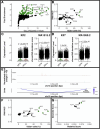
Figure 1. IL-23, a cytokine highly expressed in HPV+ cancer, reduces HPV16 E7 specific CD8 T-cell expansion and cytotoxicity. Patients with HNSCC from TCGA were stratified by known HPV status and their mRNA expression of (A) IL17A and the IL-17 polarizing factors IL6, IL1B, TGFB1, and IL23A were compared between HPV+ and HPV− HNSCC. (B) Mean expression of IL23A and IL17A mRNA of all TCGA cancers were ranked by their IL17A expression. (C) IL-23R expression of HPV-specific CD8 T-cells obtained from VRP-HPV (HPV+) and VRP-GFP (HPV−) vaccinated C3.43 tumor-bearing mice. (D) HPV16 E7-specific CD8 T-cells were incubated with 100 ng/mL of IL-23 for 5 days and characterized for HPV specificity and IFN-γ/IL-17 expression. (E) Effects of IL-23 on proliferation of HPV specific CD8 T-cells were measured through CFSE analysis, where MFI is inversely correlated to proliferation rate. (F) A cytotoxicity assay was performed for 6 hours at various effector (CD8 T-cells) to target (C3.43-gluc) ratios (E:T) and luminescence was compared between media control and IL-23 (100 ng/mL) treated CD8 T-cells. Shown are either boxplot or mean (±SEM). Functional T-cell based experiments were repeated three times with similar results. *p<0.05; **p<0.01; ***p<0.001; ****p<0.0001. HNSCC, head and neck squamous cell carcinoma; HPV, human papillomavirus; IL, interleukin; TGF, Transforming Growth Factor; CFSE, Carboxyfluorescein Succinimidyl Ester; GFP, Green Fluorescent Protein; IFN, Interferon; MFI, mean fluorescence intensity; mRNA, messenger RNA; TCGA, The Cancer Genome Atlas; VRP, Venezuelan equine encephalitis replicon particle.
Figure 2
Figure 2. Neutralizing IL-23 in HPV+ cancers reduces C3.43 tumor growth and increases HPV16 specific CD8 T-cells. (A) Experimental outline of prophylactic depletion of IL-17 and IL-23 in C57BL/6 mice bearing C3.43 HPV16+ tumors. (B) Tumor growth and (C) survival of C3.43 tumor bearing mice that continued to receive treatment with vehicle control (n=10) or either anti-IL-17 (n=10) or anti-IL-23 (n=10) antibodies dosed two times a week (50 μg/dose). (D) TILs were harvested on the day of euthanasia and analyzed by flow cytometry for HPV16 E7 tetramer+ CD8 T-cell frequency. (E) Retro-orbital bleeds were taken 14 days post-tumor challenge and analyzed for HPV16 E7 tetramer+ CD8 T-cells. (F) Other TIL frequencies measured by flow cytometry were IL-17A positive cells, (G) CD8a+ CD3+ cells, (H) CD4+ cells, and (I) T-reg cell populations. Mice received treatment until euthanasia. For flow cytometry of TILs, n ranged between 7 and 10 mice/treatment/stain. Error bars are SEM. *p<0.05; **p<0.01; ****p<0.0005; n.s., not significant. Ctrl, control; HPV, human papillomavirus; IL, interleukin; i.p., intraperitoneally; TIL, tumor-infiltrating leukocyte; T-reg, regulatory T-cells; Foxp3, forkhead box P3.
Figure 3
Figure 3. Combination of IL-23 neutralization and HPV16 E6/E7 vaccination leads to synergistic improvement of survival of C3.43 tumor-bearing mice, and improved HPV16 E7-specific CD8 T-cell tumor infiltration. (A) Schematic overview of B6 mice bearing C3.43 tumors (Day 0, n=10/group) that were treated with IL-23 neutralizing antibodies or isotype controls (Day 14–16) in combination with VRP-GFP or VRP-HPV (Day 16+23). The impact of treatment on (B) tumor growth and on (C) survival of tumor-bearing mice. TILs were analyzed throughflow cytometry to determine CD8 T-cell positive for (D) HPV E7 tetramer+ and (E) CTLA-4. (F) TIL-derived frequency of CD4 T-cells positive for CTLA-4. Error bars represent SEM. For flow cytometry, n ranged between 4 and 10 mice/treatment/stain. The experiment was repeated twice with similar results. $/#/*=p<0.05; $$/##/**=p<0.01; $$$/###/***=p<0.001; $$$$/####/****=p<0.0001. GFP, green fluorescent protein; CTLA-4, cytotoxic T-lymphocyte-associated protein 4; Ab, antibody; Ctrl, control; HPV, human papillomavirus; IL, interleukin; i.p., intraperitoneally; TIL, tumor-infiltrating leukocyte; VRP, Venezuelan equine encephalitis replicon particle.
Figure 4
Figure 4. Establishing an HPV16 E6/E7 expression tumor model. (A) Expression cassettes used to knock in HPV16 E6/E7 oncogenes or a mock control under a mouse PGK promoter and with a GFP marker gene. The mRNA expression of oncoproteins was confirmed and compared with B6MEC2, C3.43, and TC-1 by (B) qPCR normalized for GAPDH. Protein expression was confirmed by (C) western blot normalized for β-actin. (D) B6 mice were challenged with either B16-GFP (n=3) or B16-HPV (n=3) to confirm tumor take and (E) IL-23 protein was detected using Simoa, normalized for protein content per gram of tissue. *p<0.05. mRNA, messenger RNA; qPCR, quantitative PCR; GFP, green fluorescent protein; mPGK, mouse phosphoglycerate kinase; TC-1, transformed C57BL/6 1 cells; GAPDH, glyceraldehyde-3-phosphate dehydrogenase; HPV, human papillomavirus; IL, interleukin; Simoa, single molecule array;.
Figure 5
Figure 5. HPV oncoproteins drive increased IL-23 expression in macrophages. TILs isolated from B16-HPV (HPV+) and B16-GFP (HPV−) were analyzed by flow cytometry for IL-23 expression. IL-23+ cells were stratified by (A) CD45+ expression. (B) CD45+, IL-23+ immune cells were stained with (B) macrophage marker F4/80 and DC marker CD11c. (C) Gating strategy for TILs using (D) macrophage marker F4/80 and (E) the frequency of their IL-23 expression. (F) IL-23 MFI for HPV=red, GFP control=green, representing expression of IL-23 in (G) F4/80+ macrophages from either tumor model. The effect of oncoproteins expression on IL-17 expression in (H) CD45+ immune cells and (I) CD4+T-cells (Th17). Shown are mean frequencies (±SEM). Data on APCs is aggregated from three independent tumor challenge experiments with B16-GFP n=11 and B16-HPV n=12 mice. *p<0.05; **p<0.01; ****p<0.0001. APCs, antigen-presenting cells; HPV, human papillomavirus; GFP, green fluorescent protein; DC, dendritic cell; IL, interleukin; MFI, mean fluorescence intensity; TIL, tumor-infiltrating leukocyte.
Figure 6
Figure 6. HPV+ tumors express IL-23 in M2 macrophages. Macrophages and DCs were harvested from HPV− and HPV+ tumors and processed for single cell RNA+ATAC sequencing. (A) Visualization of how RNA and ATAC sequencing results are reduced into a weighted nearest neighbor analysis (WNN). (B) Cells that are Il23a RNA positive (Il23a+) are shown in yellow, cluster 0 contour is outlined in white. (C) Of the Il23a+ cells, cells that are Itgax+ (CD11c) and/or Adgre1+ (F4/80) are shown in yellow. (D) Dot plot comparing macrophage M1 (Nfkb1, Tnf, Il6, STAT1) and M2 (Mrc1, Igf1, Stab1, F13a1, Dab2) between Il23a+ and Il23a− cells. HPV, human papillomavirus; IL, interleukin; ATAC, assay for transposase-accessible chromatin; UMAP, uniform manifold approximation and projection; DCs, dendritic cells.
Figure 7
Figure 7. KLF2 and KLF7 are upregulated in TAM in an HPV-dependent manner. (A) TFs that are significantly enriched in open chromatin regions associated with Il23a expression. Candidate TFs that were more than twofold enriched compared with background open chromatin are highlighted in green. (B) Candidate TFs positively enriched in RNA level expression of Il23a+ cells (AUC) and further narrowed down by those expressed in>60% of Il23a+ cells are indicated in green. (C/D) Violin plot comparing KLF2 and KLF7 expression and motif activity score in APCs derived from HPV− and HPV+ tumors. (E) Linkage plot showing an open chromatin peak associated with Il23a expression. (F) TFs able to bind potential distal enhancer were plotted by AUC and percent expressed in Il23a+ cells (green=AUC >0.5, expression in Il23a+ cells >60%. (G) Candidate TF binding sites in the linked region were identified by scanning the sequence with their position weight matrices, using a minimum match score threshold of 95%, with relative scores>0.97 marked in green. APCs, antigen-presenting cells; AUC, area under the curve; HPV, human papillomavirus; TF, transcription factor; TAM, tumor associated macrophage.
Figure 8
Figure 8. KLF2 upregulated IL-23 in macrophages. (A) RAW264.7 mouse macrophages overexpressing KLF2 (RAWKLF2) were compared with wild-type RAW (RAWWT) for expression of KLF2 and IL-23 through qPCR. (B) Cells were grown for 72 hours, and media was analyzed for IL-23 protein concentration. Shown are mean expression values (±SEM). Data is from three biological repeats. *p<0.05; **p<0.01; ****p<0.0001. IL, interleukin; qPCR, quantitative PCR.

References

    1. Feltkamp MCW, Smits HL, Vierboom MPM, et al. Vaccination with cytotoxic T lymphocyte epitope‐containing peptide protects against a tumor induced by human papillomavirus type 16‐transformed cells. Eur J Immunol. 1993;23:2242–9. doi: 10.1002/eji.1830230929. - DOI - PubMed
    1. Smith KA, Meisenburg BL, Tam VL, et al. Lymph node-targeted immunotherapy mediates potent immunity resulting in regression of isolated or metastatic human papillomavirus-transformed tumors. Clin Cancer Res . 2009;15:6167–76. doi: 10.1158/1078-0432.CCR-09-0645. - DOI - PMC - PubMed
    1. Cassetti MC, McElhiney SP, Shahabi V, et al. Antitumor efficacy of Venezuelan equine encephalitis virus replicon particles encoding mutated HPV16 E6 and E7 genes. Vaccine (Auckl) 2004;22:520–7. doi: 10.1016/j.vaccine.2003.07.003. - DOI - PubMed
    1. Matta H, Gopalakrishnan R, Choi S, et al. Development and characterization of a novel luciferase based cytotoxicity assay. Sci Rep. 2018;8:199. doi: 10.1038/s41598-017-18606-1. - DOI - PMC - PubMed
    1. Doorbar J, Quint W, Banks L, et al. The biology and life-cycle of human papillomaviruses. Vaccine (Auckl) 2012;30 Suppl 5:F55–70. doi: 10.1016/j.vaccine.2012.06.083. - DOI - PubMed

MeSH terms

Substances

LinkOut - more resources