. 2025 Aug 19;21(1):518.
doi: 10.1186/s12917-025-04939-x.

Isolation and identification of a new porcine astrovirus 5 demonstrated that oxidative stress enhances porcine astrovirus replication

Affiliations

Isolation and identification of a new porcine astrovirus 5 demonstrated that oxidative stress enhances porcine astrovirus replication

Yiyang Du et al. BMC Vet Res. .

Abstract

Background: Porcine astrovirus (PAstV) poses a major risk to the pig industry by causing diarrhea in suckling piglets. Despite its global prevalence and five genotypes, the virus's pathogenic mechanism is not well understood due to difficulties in isolating and culturing it in vitro. Studying PAstV from clinical samples and its interaction with host cells is crucial for understanding its pathogenesis and developing antiviral treatments.

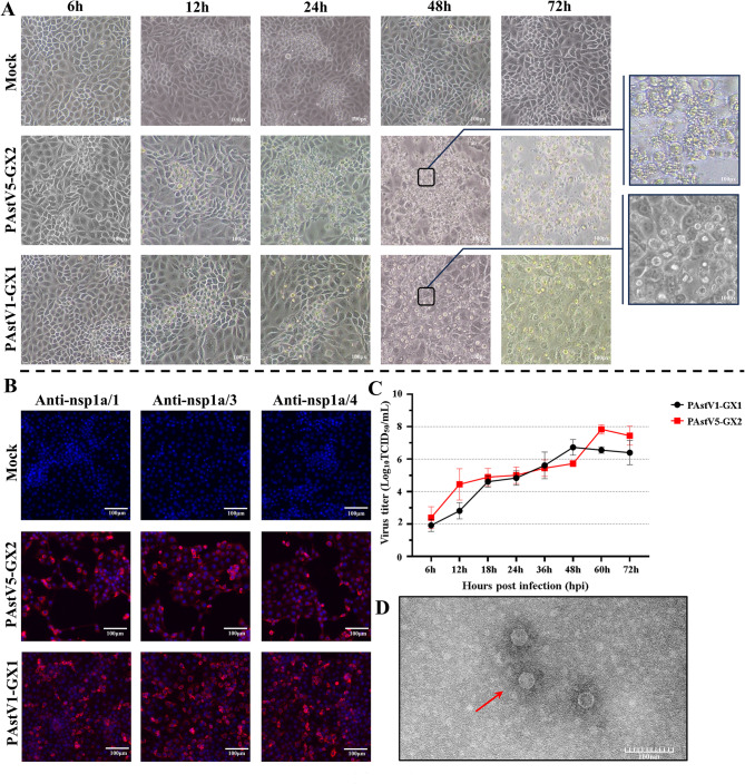
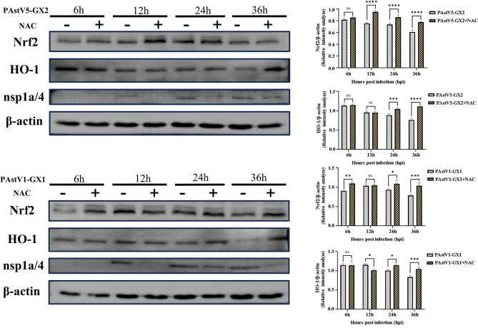
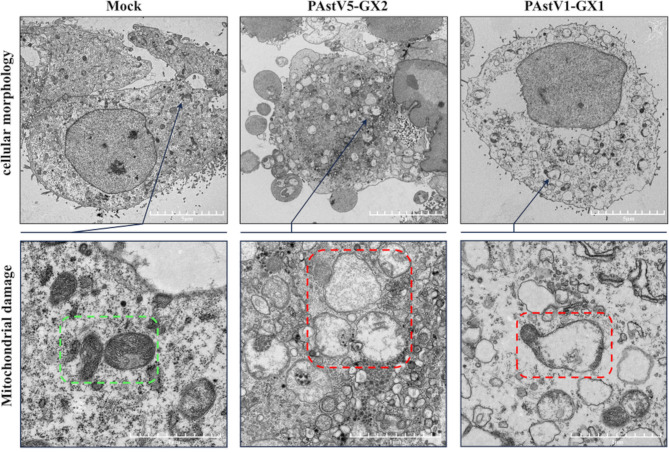
Methods: To isolate porcine astrovirus (PAstV) from clinical specimens, fecal samples from PAstV-positive pigs were collected in August 2018, inoculated into PK-15 cells, and subjected to three successive blind passages. The in vitro growth characteristics of the isolated strain were subsequently evaluated, and the morphology of the virus particles was examined through electron microscopy. The complete genome sequence of the isolated strain was determined, followed by sequence alignment, homology analysis, phylogenetic analysis, and recombination analysis. To investigate the induction of reactive oxygen species (ROS) production in PK-15 cells infected with the isolated strain, the cells were infected, and ROS production was quantified using the MitoSOX probe. Furthermore, the expression levels of the antioxidant factors Nrf2 and HO-1 were analyzed via Western blotting. Mitochondrial damage resulting from PAstV infection was observed using transmission electron microscopy, and the effect of PAstV infection on mitochondrial membrane potential was assessed using the JC-1 probe. Finally, the impact of ROS on PAstV replication was explored using IFA and RT-qPCR.

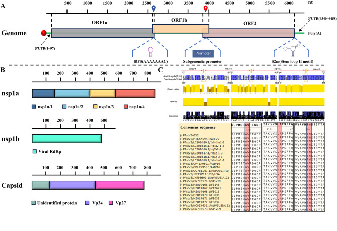
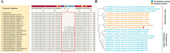
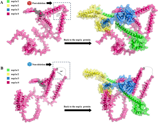
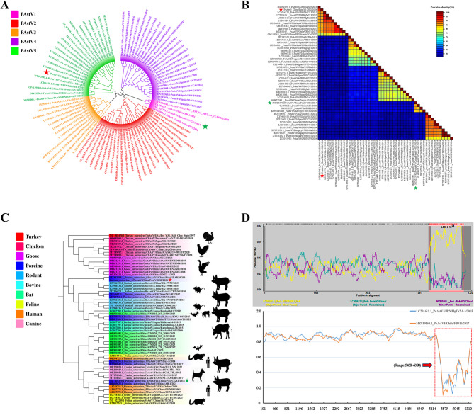
Results: In this study, a strain of PAstV was isolated from porcine fecal samples, demonstrating an ability to adapt effectively to PK-15 cells with a viral titer reaching up to 10^7.85 TCID50/mL. Genetic evolution analysis classified the isolated strain as PAstV5, revealing high genetic homology with other representative PAstV5 strains. The isolated strain was designated as PAstV5-GX2. Sequence alignment identified 11 consecutive amino acid deletions at the 3' end of ORF1a in the PAstV5-GX2 strain, resulting in alterations to the three-dimensional structure of the nsp1a/4 protein. Further investigation indicated that PAstV infection in PK-15 cells enhances mitochondrial ROS production and diminishes the protein expression levels of the antioxidant molecules Nrf2 and HO-1. Concurrently, PAstV infection induces mitochondrial swelling, cristae rupture, and vacuolization, along with a reduction in mitochondrial membrane potential. Through the application of H2O2 and NAC to modulate cellular ROS levels, it was determined that ROS can facilitate viral replication.

Conclusions and relevance: Our study successfully isolated a novel strain of PAstV5 characterized by an 11-amino acid deletion in the nsp1a protein, leading to significant alterations in the three-dimensional structure of the nsp1a/4 protein. This strain was observed to induce the production of mitochondrial ROS, downregulate the expression of Nrf2 and HO-1, and cause mitochondrial damage. Furthermore, the generation of mitochondrial ROS was found to facilitate the replication of PAstV. These findings offer valuable insights into the genetic evolution and pathogenic mechanisms of PAstV.

Keywords: Genomic analysis; Isolation; Mitochondrial damage; Oxidative stress; Porcine astrovirus 5.


Conflict of interest statement

Declarations. Ethics and consent to participate: Fecal samples in this study were collected with the consent of the piggery. Consent for publication: Not applicable. Competing interests: The authors declare no competing interests. Conflict of interest: The authors have no competing interests to declare.

Figures

Fig. 1
Fig. 1
Isolation and Characterization of PAstV5-GX2. (A) The PAstV5-GX2 strain was inoculated into PK-15 cells, and CPEs were monitored at various time intervals using microscopy, with the PAstV1-GX1 strain serving as a control. (B) PK-15 cells were subjected to either mock infection or infection with the PAstV5-GX2 and PAstV1-GX1 strains. The IFA was conducted at 36 hpi utilizing the nsp1a/1, nsp1a/3, and nsp1a/4 polyclonal antibodies as primary antibodies to detect viral replication. (C) Multi-step growth curves for the PAstV5-GX2 and PAstV1-GX1 strains were generated in PK-15 cells. (D) The morphology of PAstV5-GX2 viral particles was examined using electron microscopy
Fig. 2
Fig. 2
Whole genome sequence analysis of PAstV5-GX2. (A) Schematic diagram of the genome structure of PAstV5-GX2. (B) Schematic diagram of the PAstV5-GX2 ORF1a, ORF1b, and ORF2 encoded proteins (nsp1a, nsp1b, and capsid protein). (C) Proteolysis sites within the PAstV5-GX2 nsp1a protein were identified through comparative analysis with the nsp1a proteins of HAstV1 [21]. The precise location of the serine protease structural domain, specifically the nsp1a/3 protein, within the nsp1a polyprotein of PAstV5-GX2 was determined using the Phyre2.2 online platform. Based on the linear positioning of this structure within the entire nsp1a polyprotein, the region at the C-terminal end of the nsp1a/3 protein was designated as nsp1a/4, while the segment at the N-terminal end was designated as nsp1a/1 and nsp1a/2.Three cleavage sites in the PAstV5-GX2 nsp1a protein were highlighted using a red box
Fig. 3
Fig. 3
The PAstV5 strains originating from Asia exhibit a consecutive deletion of 11 amino acids in the nsp1a/4 protein. (A) Sequence alignment indicates that the PAstV5-GX2 strain also possesses this consecutive deletion of 11 amino acids in the nsp1a/4 protein. (B) Phylogenetic analysis demonstrates that the strains containing this deletion are exclusively derived from Asia, whereas strains lacking the deletion are predominantly found in Europe and America
Fig. 4
Fig. 4
The 3D structures of nsp1a/4 and the nsp1a protein were predicted utilizing the AlphaFold Server. Subsequently, a comparative analysis of the 3D structural differences between the PAstV5-GX2 strain (A) and the non-deletion strain JF713711.1 (B) was conducted. The spatial positions of the nsp1a/1, nsp1a/2, nsp1a/3, and nsp1a/4 proteins within the overall nsp1a protein structure are distinctly highlighted using different colors
Fig. 5
Fig. 5
Phylogenetic and recombination analyses of the PAstV5-GX2 strain. (A) A phylogenetic tree was constructed based on the complete genome sequences of PAstV, encompassing PAstV types 1 to 5. The PAstV5-GX2 and PAstV1-GX1 strains are denoted by red and green five-pointed stars, respectively. (B) An analysis of pairwise identity among the whole genome sequences of PAstV across different genotypes was performed. (C) A phylogenetic tree was constructed based on the full-length ORF2 gene of astrovirus from different species. (D) Recombinant analysis of the complete genome of PAstV5-GX2 was performed utilizing the RDP4 (upper graph) and Simplot software packages (bottom graph). A recombination event, occurring between nucleotide positions 5438 and 6388, is highlighted by a red box
Fig. 6
Fig. 6
PAstV infection resulted in oxidative stress. (A) PK-15 cells were subjected to either mock infection or infection with PAstV5-GX2 and PAstV1-GX1 at a multiplicity of infection (MOI) of 5. Mitochondrial ROS were assessed at 2, 4, 6, 8, and 12 hpi using the MitoSOX Red probe, with H2O2 treatment at 400 µM serving as a positive control. The fluorescence intensity at each time point was quantified using ImageJ software, and statistically significant differences compared to the mock-infected control were determined through three independent experiments. (B) PK-15 cells were subjected to either mock infection or infection with PAstV5-GX2 and PAstV1-GX1 at a MOI of 0.1. Cell lysates were collected at 6, 12, 24, and 36 hpi and subsequently analyzed using Western blotting to evaluate the expression levels of Nrf2, HO-1, Nsp1a-4, and β-actin. The relative expression levels of Nrf2 and HO-1, normalized to β-actin, were quantified using ImageJ software for grayscale analysis. Data shown in Graphs are presented as the mean ± SD. n = 3; ns, p > 0.05; *p < 0.05; ****p < 0.0001. These data represent the results of at least three independent experiments
Fig. 7
Fig. 7
NAC treatment effectively prevented the degradation of Nrf2 and HO-1 induced by PAstV infection. PK-15 cells were infected with either PAstV5-GX2 or PAstV1-GX1 at a MOI of 0.1, followed by either no treatment or treatment with 10 mM NAC. Cell lysates were collected at 6, 12, 24, and 36 hpi and analyzed via Western blot to evaluate the expression levels of Nrf2, HO-1, nsp1a-4, and β-actin. The relative expression levels of Nrf2 and HO-1, normalized to β-actin, were quantified using ImageJ software for grayscale analysis. Data shown in Graphs are presented as the mean ± SD. n = 3; ns, p > 0.05; *p < 0.05; **p < 0.01; ***p < 0.001; ****p < 0.0001. These data represent the results of at least three independent experiments
Fig. 8
Fig. 8
PAstV infection caused cellular and mitochondrial damage. Transmission electron microscopy was utilized to examine PK-15 cells that were either uninfected (mock) or infected with PAstV5-GX2 or PAstV1-GX1 at a MOI of 0.1, at 24 hpi. The green boxes indicate the control group displaying normal mitochondrial structures, whereas the red boxes highlight mitochondria showing signs of damage
Fig. 9
Fig. 9
PAstV infection decreased mitochondrial membrane potential. PK-15 cells were either mock-infected or infected with PAstV5-GX2 and PAstV1-GX1 at a MOI of 5. The mitochondrial membrane potential was evaluated at 2, 4, 6, 8, and 12 hpi utilizing the JC-1 probe, with CCCP treatment (10 µM) employed as a positive control. Observations were made using a fluorescence microscope at a magnification of 400x. The fluorescence intensity of each image was analyzed using ImageJ software to calculate the red/green fluorescence ratio at each time point. The bar chart illustrates the results derived from three independent experiments. Data shown in Graphs are presented as the mean ± SD. n = 3; ns, p > 0.05; ****p < 0.0001. These data represent the results of at least three independent experiments
Fig. 10
Fig. 10
Oxidative stress enhances PAstV replication. (A)PK-15 cells were pretreated with 10 mM NAC or 50 µM H2O2 for one hour prior to infection with PAstV5-GX2 and PAstV1-GX1 at a MOI of 0.1, respectively. Following infection, NAC and H2O2 were continuously supplemented in the cell culture medium. At 12, 24, and 36 hpi, the cells were analyzed for mtROS using the MitoSOX Red probe, indicated by red fluorescence, or fixed for IFA to evaluate viral protein levels using a polyclonal antibody against nsp1a/4, indicated by green fluorescence. (B)Simultaneously, cells and supernatants from different treatment groups were collected for total RNA extraction, and virus copy numbers of PAstV5-GX2 and PAstV1-GX1 were measured using RT-qPCR. Data shown in Graphs are presented as the mean ± SD. n = 3; ns, p > 0.05; **p < 0.01; ***p < 0.001; ****p < 0.0001. These data represent the results of at least three independent experiments

Similar articles

References

    1. Wohlgemuth N, Honce R, Schultz-Cherry S. Astrovirus evolution and emergence. Infect Genet Evol. 2019;69:30–7. - PMC - PubMed
    1. van Hemert FJ, Lukashov VV, Berkhout B. Different rates of (non-)synonymous mutations in astrovirus genes; correlation with gene function. Virol J. 2007;4:25. - PMC - PubMed
    1. Ykema M, Tao YJ. Structural insights into the human astrovirus capsid. Viruses. 2021;13(5):821. - PMC - PubMed
    1. Lulla V, Firth AE. A hidden gene in astroviruses encodes a Viroporin. Nat Commun. 2020;11(1):4070. - PMC - PubMed
    1. Du Y, Ji C, Liu T, Zhang W, Fang Q, Dong Q, Li M, Wang H, Chen Y, Ouyang K, et al. Identification of a novel protein in Porcine astrovirus that is important for virus replication. Vet Microbiol. 2021;255:108984. - PubMed

Substances

LinkOut - more resources