

Protective effect of ammonium trichloride tellurate (AS101) on ovarian injury induced by chemotherapy drug doxorubicin in rats
- PMID: 40830524
- PMCID: PMC12363043
- DOI: 10.1186/s13048-025-01709-z
Protective effect of ammonium trichloride tellurate (AS101) on ovarian injury induced by chemotherapy drug doxorubicin in rats
Abstract
Objective: To investigate the protective effect of ammonium trichloride tellurate (AS101) on doxorubicin induced ovarian function damage in rats.
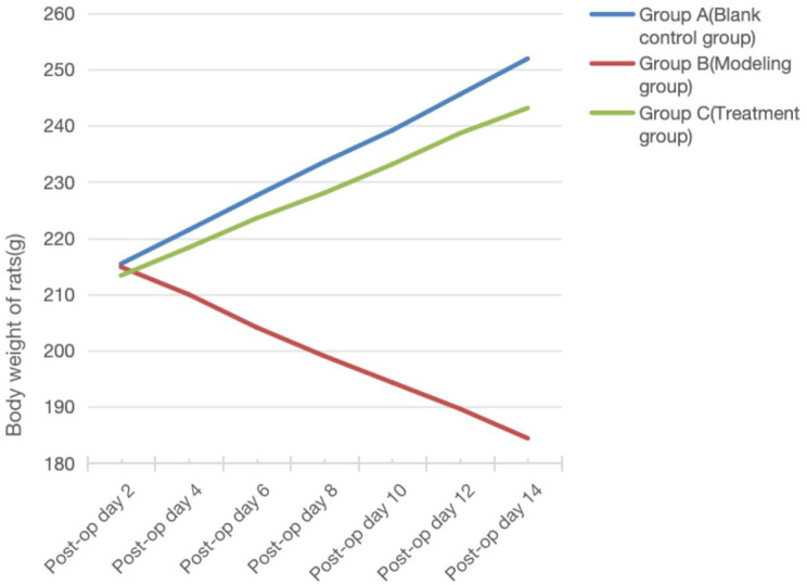
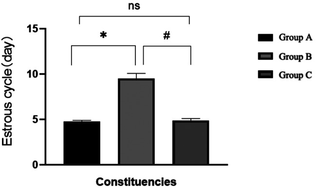
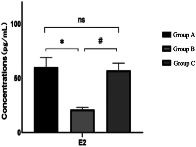
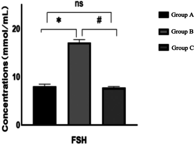
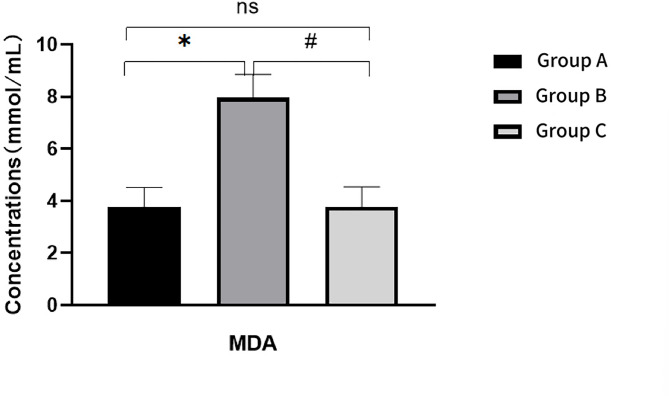
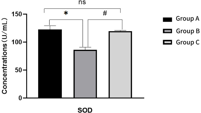
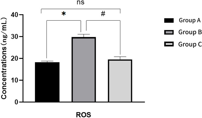
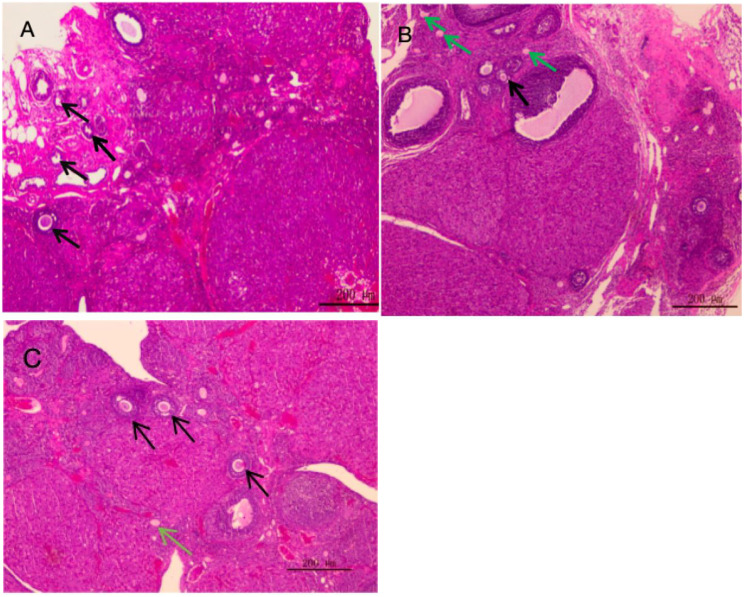
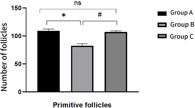
Methods: Eighteen female SD rats were randomly divided into three groups with 6 rats in each group: group A blank control group (0.9% normal saline 0.1 ml intraperitoneally for 21 consecutive days), group B (0.9% normal saline 0.1 ml intraperitoneally for 5 days, doxorubicin 10 mg / kg intraperitoneally on day 6, and continued to be injected with normal saline for 15 days), group C (0.9% normal saline 0.1 ml intraperitoneally for 5 days, doxorubicin 10 mg / kg intraperitoneally on day 6, and continued to be injected with AS101 twice every other day). The contents to be recorded by the operator team include: (1) before operation: ① observe the general state of rats in each group and record the weight of rats. ② Vaginal smears of rats in each group were observed at the same time every day to observe the estrous cycle of rats. (2) After operation: ① observe the general state of rats in each group, read the weight of rats, and record the data Vaginal smears of rats in each group were observed at the same time every day after operation to observe the estrous cycle of rats. ③ At the end of the postoperative observation period, blood samples were taken from the tail of rats to measure the serum levels of estrogen 2 (E2), follicle stimulating hormone (FSH), and anti-M ü llerian hormone (AMH). ④ At the end of the observation period, the animals were sacrificed to observe the development of bilateral ovarian follicles. ⑤ At the end of the observation period, blood was taken from the abdominal aorta of rats after anesthesia, and the upper serum was separated after centrifugation at 3000 rpm. The levels of malondialdehyde (MDA), superoxide dismutase (SOD) and reactive oxygen species (ROS) were measured according to the operation methods of the kit instructions.
Result: The body weight of rats in group B was significantly lower than that in group A, while that in group C was significantly higher than that in group B (P < 0.05). The estrous cycle of rats in group B was disordered, and the estrous cycle of rats in group C was restored regularly (P < 0.05). The serum estradiol(E2) and AMH levels in group C were significantly higher than those in group B, and the FSH level was significantly lower than that in group B. Compared with group B, the number of primary follicles increased and the number of atretic follicles decreased in group C (P < 0.05). The SOD level in group B was lower than that in group A, and that in group C was higher than that in group B (P < 0.05). The levels of MDA and ROS in group B were higher than those in group A (P < 0.05), while those in group C were lower than those in group B (P < 0.05).
Conclusion: AS101 can protect the damage of doxorubicin on ovarian function in rats, and its mechanism is related to its antioxidant effect.
Clinical trial number: Not applicable.
Keywords: AS101; Chemotherapy; Ovarian; Oxidative stress; Reproductive endocrine.
© 2025. The Author(s).
Conflict of interest statement
Declarations. Competing interests: The authors declare no competing interests.
Figures


References
-
- Siegel RL, Miller KD, Wagle NS, Jemal A. Cancer statistics, 2023. CA Cancer J Clin. 2023;73(1):17–48. - PubMed
MeSH terms
Substances
Grants and funding
- 361007/Hebei Province Government Funded Clinical Medical Excellence Project (Leader)
- ZD202314/Science and Technology Project of Changzhou Health Commission
- 2022CZBJ074/Top Talent of Changzhou "The 14th Five-Year Plan" High-Level Health Talents Training Project
- F202138/the Maternal and Child Health Research Project of Jiangsu Province
LinkOut - more resources
Full Text Sources
