. 2025 Aug 19;10(1):196.
doi: 10.1038/s41541-025-01254-2.

Stabilization of norovirus GII.3 virus-like particles by rational disulfide engineering

Affiliations

Stabilization of norovirus GII.3 virus-like particles by rational disulfide engineering

Christopher Warren et al. NPJ Vaccines. .

Abstract

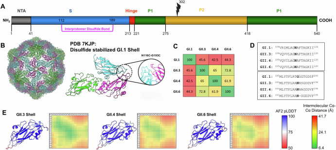
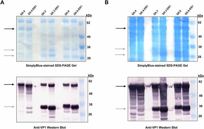
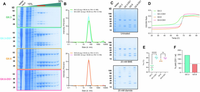
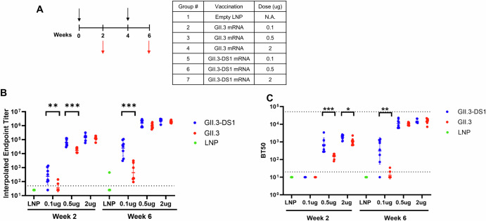
Noroviruses are non-enveloped, single-stranded positive-sense RNA viruses and the leading cause of gastroenteritis worldwide. The major capsid protein, VP1, can self-assemble into non-infectious virus-like particles (VLPs), representing an attractive vaccine platform. It was demonstrated that engineered disulfide bonds within VP1 could significantly stabilize VLPs of the archetypal GI.1 strain. Here, we apply a similar strategy to VLPs of multiple circulating GII genotypes. We find that engineered disulfide mutations can significantly stabilize VLPs of the GII.3 strain, but not the closely related GII.6 strain. Disulfide-stabilized GII.3 VLPs (GII.3-DS1) exhibit increased yields, greater homogeneity, and higher thermal stability compared to wild-type GII.3 VLPs. GII.3-DS1 VLPs are a superior reagent in immunological assays compared to the wild-type counterpart. Importantly, mRNA encoding GII.3-DS1 elicits superior humoral immune responses compared to wild-type GII.3 mRNA in mice. These results demonstrate the utility of rational VLP stabilization for advancing vaccine development efforts.


Conflict of interest statement

Competing interests: All co-authors are employees of Merck Sharp & Dohme LLC, a subsidiary of Merck & Co., Inc., Rahway, NJ, USA and may own stock or hold stock options in Merck & Co., Inc., Rahway, NJ, USA. This does not alter the authors’ adherence to all journal policies on competing interests and sharing data and materials. In addition, a subset of the authors is listed as inventors on a patent surrounding this work held by Merck & Co., Inc., Rahway, NJ, USA.

Figures

Fig. 1
Fig. 1. Structure-guided stabilization of norovirus VLPs.
A Domain map of the norovirus VP1 protein showing the shell (S), hinge, and protruding (P1/P2) domains. Protease cleavage site present in GII.3 and GII.6 shown at residue 302 (lightning bolt). Engineered disulfide bond (DS1) between interprotomer residues N112 and N189 shown in purple. B Structure of the disulfide-stabilized GI.1 shell (PDB 7KJP) colored by chain. Left shows the intact T = 3 VLP. Asymmetric unit shown on the right with the interprotomer disulfide highlighted. C Sequence identity matrix of full-length VP1 based on the alignment of 4 norovirus genotypes. D Sequence alignment of VP1 from 4 norovirus genotypes in the regions of the engineered disulfide bond. Cysteine mutation positions are in bold. E Alphafold models of the shell domains of three GII genotypes colored by pLDDT. Monomeric models were aligned to the GI.1 T = 3 asymmetric unit (PDB 7KJP) and their energies were minimized. Interprotomer Ca-Ca distance matrices were calculated between loops containing residues 105–120 (y-axis) and 181–196 (x-axis) to predict proximity of residue pairs within the T = 3 VLP structure.
Fig. 2
Fig. 2. VP1 protein is expressed successfully from transiently transfected Expi293 cells.
Cells were transfected with expression plasmids encoding various strains of norovirus VP1. Clarified supernatant (2.6 mg/lane, A) and lysate (15 mg/lane, B) fractions were separated via SDS-PAGE and either stained with SimplyBlue Safestain (upper panels) or blotted onto membranes and probed with an anti-VP1 antibody (lower panels). The position of full-length VP1 is denoted by black arrows. The positions of cleaved fragments of strains GII.3 and GII.6 are denoted by gray arrows. Marker proteins in kilodaltons (kDa) are indicated.
Fig. 3
Fig. 3. DS1 mutation stabilizes GII.3 VLPs.
A Sucrose gradient purification of GII.3 and GII.6 VLPs with and without DS1 mutation. Fractions 5–7 (red line) were pooled for downstream analyses. Gradient input denoted input. B Dynamic light scattering (DLS) of GII.3 and GII.6 VLPs. Z-average (z-avg) and polydispersity index (PdI) values are shown in the insert. C SDS-PAGE analysis of GII.3 and GII.6 VLPs in either the untreated condition (top) or after treatment with 20 mM BME (middle) or 20 mM diamide (bottom) for 1 h at room temperature. D nanoDSF measurements of GII.3 and GII.6 VLPs. The line represents the average of 3 technical replicates. E Quantification of melting temperature (Tm) from nanoDSF experiments, showing a significant increase in thermal stability of GII.3 VLPs upon introduction of the DS1 mutation. Significant values determined by unpaired t-test (p < 0.0001). F Relative yields of DS1 mutant compared to wild-type after sucrose gradient purification of VLPs.
Fig. 4
Fig. 4. DS1 mutation leads to more uniform VLPs.
Representative negative-stain electron micrographs (nsEM) and 2D classes of GII.3 (A), GII.3-DS1 (B), GII.6 (C), and GII.6-DS1 (D) VLPs. 2D class averages of intact VLPs are in the panel set on the right. nsEM magnification is 58,000X. VLPs were stored frozen at -80⁰C until processed for imaging. A GII.3 2D class averages derived from 139 particles picked from 27 micrographs. B GII.3-DS1 2D class averages derived from 97 particles picked from 26 micrographs. C GII.6 2D class averages derived from 205 particles picked from 27 micrographs. D GII.6-DS1 2D class averages derived from 314 particles picked from 24 micrographs.
Fig. 5
Fig. 5. GII.3 and GII.3-DS1 VLPs bind to human saliva samples and monoclonal antibodies.
A ELISA plates were coated with 51 human saliva samples and incubated with GII.3 or GII.3-DS1 VLPs at 20 µg/mL. Bound VLPs were detected using polyclonal sera against GII.3. Relative luminescent units (RLU) of GII.3 and GII.3-DS1 are reported on the x- and y-axes, respectively. Data are fitted into a simple linear regression model using GraphPad Prism software. B Plates were coated with 50 ng of either GII.3 or GII.3-DS1 VLP and incubated with 19 unique human monoclonal antibodies starting at 4 µg/mL and diluted in a four-fold, ten-point dilution series. Bound antibodies were detected using goat anti-human Ig-Fc fragment conjugated to horseradish peroxidase (HRP). Interpolated endpoint titer for each antibody against either GII.3 or GII.3-DS1 is reported on the x- and y-axes, respectively. Data are fitted into a simple linear regression model using GraphPad Prism software.
Fig. 6
Fig. 6. Mouse immunogenicity study of mRNAs encoding either GII.3 or GII.3-DS1, using assay reagent GII.3-DS1 VLP.
A In vivo immunogenicity study design. BALB/c mice (N = 8 per group) were immunized with 0.1, 0.5, or 2 µg of mRNA at weeks 0 and 4 (black arrows). An empty lipid nanoparticle (LNP) was immunized as a negative control. Blood was drawn at weeks 2 and 6 (red arrows). Serum antibody ELISA titers (B) and HBGA blockade antibody titers (C) against GII.3-DS1 VLP. Serum of mice immunized with mRNAs encoding either GII.3 (red dots) or GII.3-DS1 (blue dots) was assayed. B, C Limits of detection are indicated with horizontal dotted lines (lower limit titer = 50 in (B) and 20 in (C), upper limit titer = 51,200 in (C). Geometric mean titers with geometric standard deviation are shown in a scatter dot plot. P values were determined using GraphPad Prism software (p < 0.05*, p < 0.01**, p < 0.001***).

References

    1. Lopman, B. A., Steele, D., Kirkwood, C. D. & Parashar, U. D. The vast and varied global burden of norovirus: prospects for prevention and control. PLoS Med.13, e1001999 (2016). - PMC - PubMed
    1. Teunis, P. F. et al. Norwalk virus: how infectious is it?. J. Med. Virol.80, 1468–1476 (2008). - PubMed
    1. de Graaf, M., van Beek, J. & Koopmans, M. P. Human norovirus transmission and evolution in a changing world. Nat. Rev. Microbiol.14, 421–433 (2016). - PubMed
    1. Verhoef, L. et al. Norovirus genotype profiles associated with foodborne transmission, 1999–2012. Emerg. Infect. Dis.21, 592–599 (2015). - PMC - PubMed
    1. Bartsch, S. M., Lopman, B. A., Ozawa, S., Hall, A. J. & Lee, B. Y. Global economic burden of norovirus gastroenteritis. PLoS One11, e0151219 (2016). - PMC - PubMed

LinkOut - more resources