Protective and therapeutic potentials of Trichinella spiralis larval antigen in murine induced colitis
- PMID: 40830617
- PMCID: PMC12365207
- DOI: 10.1038/s41598-025-13229-3
Protective and therapeutic potentials of Trichinella spiralis larval antigen in murine induced colitis
Abstract
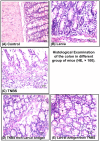
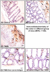
The intolerable side effects and clinical limitations of current conventional therapies for inflammatory bowel diseases (IBDs), there is a pressing need for alternative treatment options. Helminthes adapt immune responses of their hosts to reduce immune-mediated IBDs. The identification of the mechanism responsible for this beneficial effect on IBDs will provide another feasible approach to treating these diseases. The study was designed to investigate the possible protective and therapeutic role of Trichinella spiralis (T. spiralis) crude larval antigen extract in mice challenged with 2,4,6-trinitrobenzene sulfonic acid (TNBS) to induce colitis. Colitis was induced by intra-colonic instillation of TNBS (5 mg/ml in 50% ethanol), preceded or followed by intra-peritoneal (i.p.) administration of a single dose of T. spiralis crude larval antigen extract (100 µg/mouse). Colonic damage was assessed macroscopically and microscopically, and the expression of myeloperoxidase (MPO) was evaluated by immunohistochemistry. Colonic interleukin-10 (IL-10) and serum nitric oxide (NO) levels were also measured. Administration of T. spiralis crude larval antigen extract before induction of colitis reduced colitis severity as demonstrated by reduced colon weight-to-length ratio, improved macroscopic and microscopic scores, increased colonic IL-10 expression, and diminished colonic MPO protein expression. Moreover, there was a significant negative correlation between serum NO and colonic IL-10 levels. In addition, the preventive potential of T. spiralis crude larval antigen extract against TNBS-induced colitis was more prominent than its therapeutic effect. These findings support the hypothesis that T. spiralis has both prophylactic and therapeutic potential in inflammatory bowel diseases, which may be via an increase in IL-10 with predominance of its prophylactic role.
Keywords: Trichinella spiralis; Colitis; IL-10 and myeloperoxidase; NO; TNBS.
© 2025. The Author(s).
Conflict of interest statement
Declarations. Competing interests: The authors declare no competing interests. Ethics approval and consent to participate: The study protocol was approved by the ethical committee of the Faculty of Medicine, Assiut University (IRB code:17300218).
Figures


Similar articles
-
Trichinella spiralis adult excretory-secretory antigen promotes peripheral regulatory T cell differentiation and attenuates experimental colitis via TGF-β-like mechanisms.Parasit Vectors. 2025 Jul 1;18(1):240. doi: 10.1186/s13071-025-06877-x. Parasit Vectors. 2025. PMID: 40597393 Free PMC article.
-
Prescription of Controlled Substances: Benefits and Risks.2025 Jul 6. In: StatPearls [Internet]. Treasure Island (FL): StatPearls Publishing; 2025 Jan–. 2025 Jul 6. In: StatPearls [Internet]. Treasure Island (FL): StatPearls Publishing; 2025 Jan–. PMID: 30726003 Free Books & Documents.
-
The Black Book of Psychotropic Dosing and Monitoring.Psychopharmacol Bull. 2024 Jul 8;54(3):8-59. Psychopharmacol Bull. 2024. PMID: 38993656 Free PMC article. Review.
-
Evaluating the Anti-inflammatory Efficacy of a Novel Bipyrazole Derivative in Alleviating Symptoms of Experimental Colitis.Curr Mol Pharmacol. 2024;17:e18761429333261. doi: 10.2174/0118761429333261241021034043. Curr Mol Pharmacol. 2024. PMID: 39604158
-
Oral budesonide for induction of remission in ulcerative colitis.Cochrane Database Syst Rev. 2015 Oct 26;2015(10):CD007698. doi: 10.1002/14651858.CD007698.pub3. Cochrane Database Syst Rev. 2015. PMID: 26497719 Free PMC article.
References
-
- De Souza, H. S. P., Fiocchi, C. & Iliopoulos, D. The IBD interactome: an integrated view of aetiology, pathogenesis and therapy. Nat. Rev. Gastroenterol. Hepatol.14, 739–749 (2017). - PubMed
MeSH terms
Substances
LinkOut - more resources
Full Text Sources
Research Materials
Miscellaneous

