

Mechanism of bactericidal efficacy against nosocomial pathogenic Staphylococcus aureus strain caused by fatty acids from Hermetia illucens larvae fat
- PMID: 40830635
- PMCID: PMC12365018
- DOI: 10.1038/s41598-025-15858-0
Mechanism of bactericidal efficacy against nosocomial pathogenic Staphylococcus aureus strain caused by fatty acids from Hermetia illucens larvae fat
Abstract
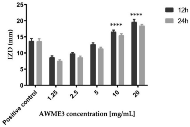
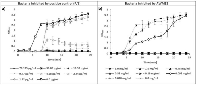
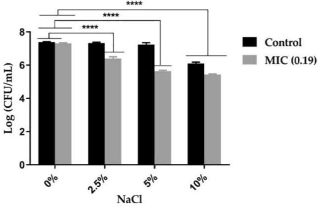
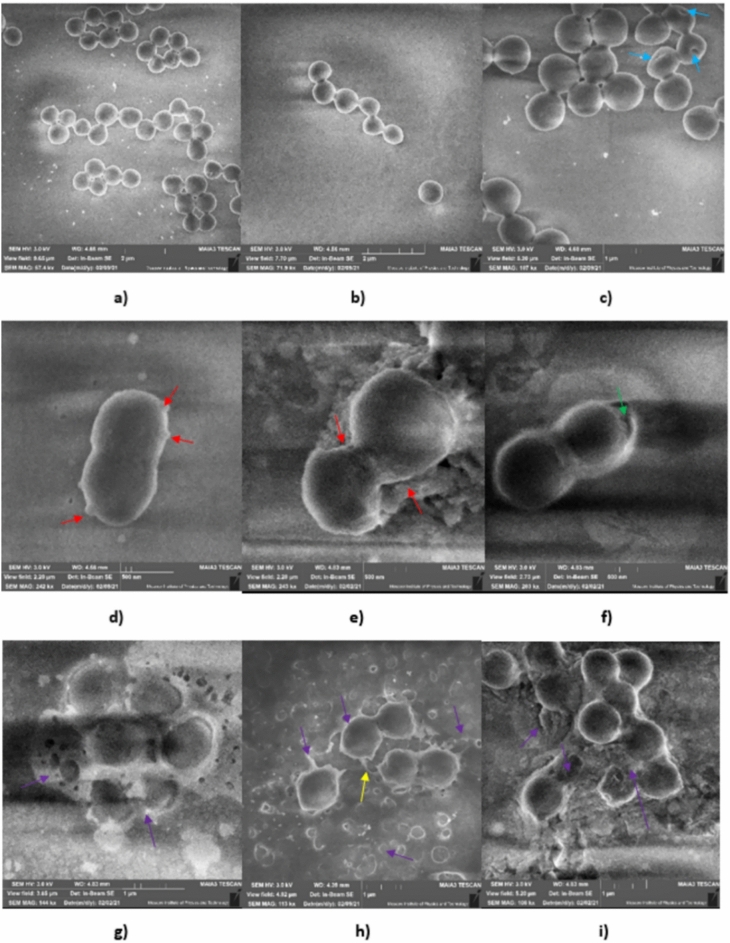
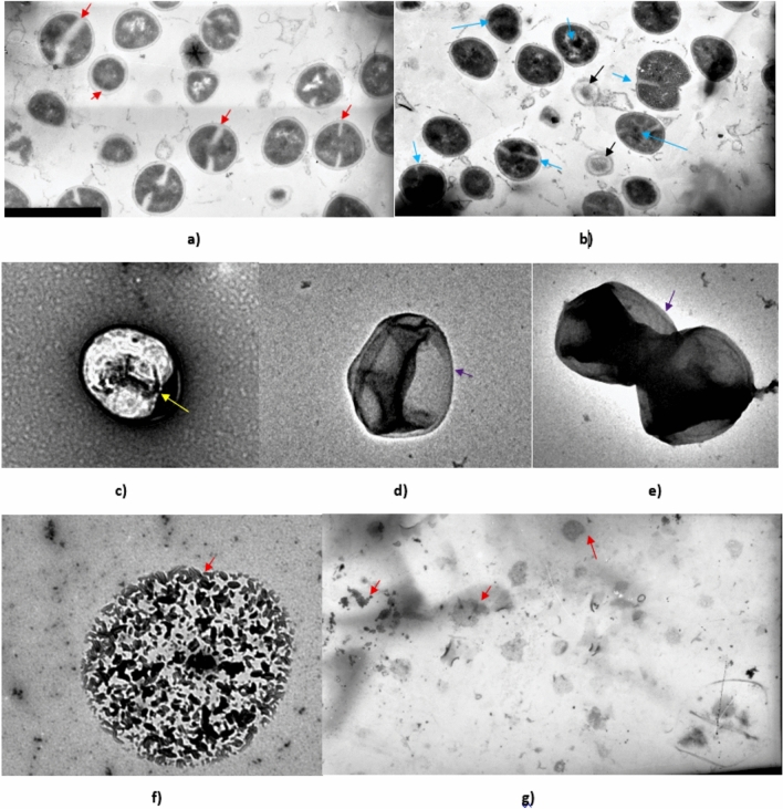
Hermetia illucens (HI) is a promising insect that widely employed as a sustainable source of food and has been recently used as a successful antimicrobial agent. Fatty acids extracted sequentially from HI larvae fight against MDR nosocomial pathogenic bacteria such as Staphylococcus aureus. This strain is resistant to various antibiotics, causing many issues and deaths in healthcare sectors. The present study aimed to elucidate the mechanism of bactericidal efficacy of fatty acids (FAs) in HI larvae fat against S. aureus ATCC 55804 strain. The disk diffusion assay, minimum inhibitory concentration (MIC), minimum bactericidal concentration (MBC), and half of the minimum inhibitory concentration (MIC50) applied in this study, proved the antimicrobial activity of fatty acids. The mechanism of FAs action was evaluated by several approaches, including inhibition of the bacterial growth curves and salt tolerance assays, scanning electron (SEM) and transmission electron (TEM) microscopies. S. aureus ATCC 55804 was resistant to 30% out of ten tested antibiotics belonging to different classes. In addition, microscopic observations showed the inhibitory effect of acidic water methanol extract (AWME3) by targeting of the S. aureus ATCC 55804 cell membrane and causing the considerable morphological alterations on the bacterial wall and destruction its cytoplasmic contents leading to the cellular content release and cell death. This study revealed the potential efficacy of AWME3 as a novel therapeutic antibacterial agent effective against resistant nosocomial bacterial pathogens.
Keywords: Hermetia illucens; AWME3 extract Fatty acids; MDR bacteria; SEM; Salt tolerance; TEM.
© 2025. The Author(s).
Conflict of interest statement
Declarations. Competing interests: The authors declare no competing interests. Human and animal resources: The authors state that no humans or animals were involved in the study.
Figures


Similar articles
-
Bacterial Outer Membrane Permeability Increase Underlies the Bactericidal Effect of Fatty Acids From Hermetia illucens (Black Soldier Fly) Larvae Fat Against Hypermucoviscous Isolates of Klebsiella pneumoniae.Front Microbiol. 2022 May 6;13:844811. doi: 10.3389/fmicb.2022.844811. eCollection 2022. Front Microbiol. 2022. PMID: 35602017 Free PMC article.
-
The antibacterial activity and therapeutic potential of the amphibian-derived peptide TB_KKG6K.mSphere. 2025 Jun 25;10(6):e0101624. doi: 10.1128/msphere.01016-24. Epub 2025 May 19. mSphere. 2025. PMID: 40387366 Free PMC article.
-
Repurposing Etalocib suppresses multidrug-resistant Staphylococcus aureus by disrupting the bacterial membrane.BMC Microbiol. 2025 Aug 1;25(1):472. doi: 10.1186/s12866-025-04163-5. BMC Microbiol. 2025. PMID: 40750851 Free PMC article.
-
A systematic review and meta-analysis revealed a high-level antibiotic resistance of bovine mastitis Staphylococcus aureus in Iran.Res Vet Sci. 2023 Aug;161:23-30. doi: 10.1016/j.rvsc.2023.05.016. Epub 2023 Jun 5. Res Vet Sci. 2023. PMID: 37302281
-
Applications of Lab on a Chip in Antimicrobial Susceptibility of Staphylococcus aureus: A Systematic Review.Medicina (Kaunas). 2023 Sep 26;59(10):1719. doi: 10.3390/medicina59101719. Medicina (Kaunas). 2023. PMID: 37893437 Free PMC article.
References
-
- Foster, T. J. Antibiotic resistance in Staphylococcus aureus. Current status and future prospects. FEMS Microbiol. Rev.41, 430–449 (2017). - PubMed
MeSH terms
Substances
Grants and funding
LinkOut - more resources
Full Text Sources
Medical
Research Materials
Miscellaneous
