

Radiomics-based classification of pediatric dental trauma in periapical radiographs: a preliminary study
- PMID: 40830854
- PMCID: PMC12362901
- DOI: 10.1186/s12880-025-01877-w
Radiomics-based classification of pediatric dental trauma in periapical radiographs: a preliminary study
Abstract
Purpose: To evaluate the feasibility and effectiveness of radiomic analysis of the periapical region on periapical radiographs for classifying the subtypes of dental trauma in pediatric patients.
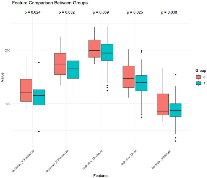
Methods: A retrospective analysis was conducted on 111 pediatric patients who presented with dental trauma and underwent periapical radiography. Patients were categorized into tooth concussion (n = 23) and tooth fracture (n = 88) groups on the basis of the type of injury. Patients were randomly stratified into training (n = 78; concussion: 16, fracture: 62) and testing (n = 33; concussion: 7, fracture: 26) cohorts at a 7:3 ratio. Regions of interest were manually delineated around the apical foramen, and radiomic features were subsequently extracted. Feature selection was performed using the intraclass correlation coefficient, the Pearson correlation coefficient, and one-way ANOVA. A support vector machine classifier was constructed based on the selected features. The performance of the radiomic model was evaluated using the area under the receiver operating characteristic curve (AUC), sensitivity, and specificity.
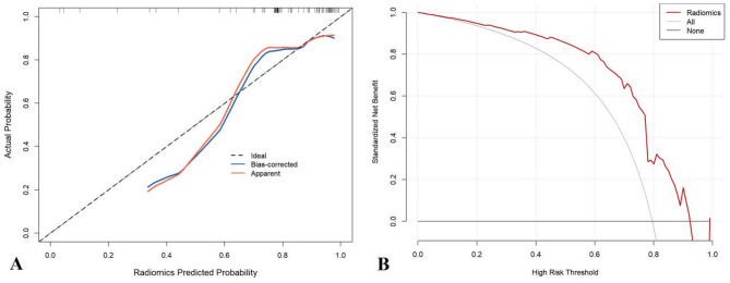
Results: A total of 21 radiomic features were selected to construct the final model. In the training cohort, the model achieved an AUC of 0.766 (95% confidence interval (CI): 0.605–0.927), with a sensitivity of 1.000 and a specificity of 0.500 in differentiating the subtypes of dental trauma. In the testing cohort, the model yielded an AUC of 0.758 (95% CI: 0.538–0.979), with a sensitivity of 0.808 and a specificity of 0.714.
Conclusion: Radiomic analysis of periapical radiographs shows promise in distinguishing between tooth concussion and fracture in pediatric patients. Further validation is needed to confirm its clinical utility and broader applicability.
Supplementary Information: The online version contains supplementary material available at 10.1186/s12880-025-01877-w.
Keywords: Children; Dental trauma; Periapical radiography; Radiomics.
Conflict of interest statement
Declarations. Ethics approval and consent to participate: This study was conducted in accordance with the Declaration of Helsinki and was approved by the Institutional Review Board of Deyang Stomatological Hospital. Patient informed consent was waived. Consent for publication: Not applicable. Competing interests: The authors declare no competing interests.
Figures


Similar articles
-
Assessment of unfilled and filled canals in mandibular incisors: comparison between periapical radiography and cone-beam computed tomography.Odontology. 2025 Aug 14. doi: 10.1007/s10266-025-01174-6. Online ahead of print. Odontology. 2025. PMID: 40813553
-
Comparison between periapical radiography and cone beam computed tomography for the diagnosis of anterior maxillary trauma in children and adolescents.Dent Traumatol. 2022 Feb;38(1):62-70. doi: 10.1111/edt.12706. Epub 2021 Jul 18. Dent Traumatol. 2022. PMID: 34275178
-
The effect of adenoid hypertrophy on growth-development level and dental maturation: a 15-year retrospective radiographs study.BMC Oral Health. 2025 Jul 28;25(1):1266. doi: 10.1186/s12903-025-06600-3. BMC Oral Health. 2025. PMID: 40721764 Free PMC article.
-
Panoramic radiography in dental diagnostics.Swed Dent J Suppl. 1996;119:1-26. Swed Dent J Suppl. 1996. PMID: 8971997 Review.
-
Diagnostic Accuracy of Cone-beam Computed Tomography and Conventional Radiography on Apical Periodontitis: A Systematic Review and Meta-analysis.J Endod. 2016 Mar;42(3):356-64. doi: 10.1016/j.joen.2015.12.015. J Endod. 2016. PMID: 26902914
References
Grants and funding
LinkOut - more resources
Full Text Sources
