[Preprint]. 2025 Aug 12:2025.08.08.669392.
doi: 10.1101/2025.08.08.669392.

A non-catalytic role for RFC in PCNA-mediated processive DNA synthesis

A non-catalytic role for RFC in PCNA-mediated processive DNA synthesis

Gabriella N L Chua et al. bioRxiv. .

Abstract

The ring-shaped sliding clamp PCNA enables DNA polymerases to perform processive DNA synthesis during replication and repair. The loading of PCNA onto DNA is catalyzed by the ATPase clamp loader RFC. Using a single-molecule platform to visualize the dynamic interplay between PCNA and RFC on DNA, we unexpectedly discovered that RFC continues to associate with PCNA after loading, contrary to the conventional view. Functionally, this clamp-loader/clamp complex is required for processive DNA synthesis by polymerase δ (Polδ), as the PCNA-Polδ assembly is inherently unstable. This architectural role of RFC is dependent on the BRCT domain of Rfc1, and mutation of its DNA-binding residues causes sensitivity to DNA damage in vivo. We further showed the FEN1 flap endonuclease can also stabilize the PCNA-Polδ interaction and mediate robust synthesis. Overall, our work revealed that, beyond their canonical enzymatic functions, PCNA-binding proteins harbor non-catalytic functions essential for DNA replication and genome maintenance.


Conflict of interest statement

DECLARATION OF INTERESTS

The authors declare no competing interests.

Figures

Figure 1.
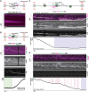
Figure 1.. Clamp-loader/clamp complexes bind and slide on dsDNA.
(A) Schematic of the experimental setup. A single DNA molecule containing a 3-kb ssDNA gap flanked by 10-kb and 6-kb dsDNA arms was tethered between a pair of optically trapped beads through biotin-streptavidin linkage. The tether was moved to a channel containing LD655-PCNA, Cy3-RFC, RPA, and ATP, and their behavior was imaged via dual-color scanning confocal fluorescence microscopy. (B) Two representative kymographs showing gapped DNA tethers incubated with 8 nM LD655-PCNA, 8 nM Cy3-RFC, and 100 nM RPA. The red laser was turned off briefly to confirm the presence of the RFC fluorescence signal. Arrows denote the events when PCNA is engaged with the 3’ recessed end of the ssDNA gap. (C) Dwell times of PCNA binding events at the 3’ recessed end of the ssDNA gap. Bars represent mean and SEM (N = 61 events). See also Figures S1 and S2.
Figure 2.
Figure 2.. Clamp-loader/clamp complexes can be topologically bound to DNA.
(A) Representative kymograph showing a double-stranded λ DNA tether incubated with 8 nM LD655-PCNA, 8 nM Cy3-RFC, and 100 nM RPA with ATP. Red laser was turned off briefly to confirm the presence of green RFC signals. (B) Kymograph showing a ds λ DNA tether first incubated in a channel containing 8 nM LD655-PCNA, 8 nM Cy3-RFC, and 100 nM RPA with ATP and then moved into a separate channel containing a high-salt buffer (500 mM NaCl) with ATP. (C) Fraction of clamp-loader/clamp complexes (CLCs) that remained bound to or fell off dsDNA when challenged with high salt (N = 54 from 17 independent tethers). See also Figure S2.
Figure 3.
Figure 3.. The BRCT domain of RFC promotes CLC formation on DNA.
(A) Domain structure of Rfc1 and Rfc1ΔBRCT. The BRCT domain is highlighted in green. (B) Two representative kymographs showing a gapped DNA tether incubated with 8 nM LD655-PCNA, 8 nM LD555-RFCΔBRCT, and 100 nM RPA. (C) Fraction of trajectories on gapped DNA containing CLCs or PCNA only when RFC (N = 46 from 6 independent tethers) or RFCΔBRCT (N = 33 from 8 independent tethers) was used. (D) Diffusion coefficients (D) for PCNA trajectories sliding on DNA when RFC (N = 22 from 5 independent tethers) or RFCΔBRCT (N = 6 from 6 independent tethers) was used. Bars represent mean and SEM. Significance was calculated using a two-tailed unpaired t-test with Welch’s correction. See also Figures S1 and S3.
Figure 4.
Figure 4.. Clamp-loader/clamp complexes assemble with Polδ to support DNA synthesis.
(A) (Top) Schematic of experimental setup. A single gapped DNA molecule was tethered between a pair of optically trapped beads held at a constant distance. The tether was moved to a channel containing Polδ, PCNA, RFC, and AF488-RPA with ATP and dNTPs. The behavior of RPA was imaged using scanning confocal fluorescence microscopy. (Bottom) Kymograph showing RPA clearing during fill-in of the ssDNA gap when incubated with 20 nM Polδ, 5 nM PCNA, 5 nM RFC, and 20 nM AF488-RPA. (B) (Top) Schematic of experimental setup where the distance between beads was held constant starting at a force of 9 pN, and fill-in was monitored by an increase in the force as the tether shortened. (Bottom) Kymograph and associated force changes showing fill-in of a gapped DNA tether incubated with 20 nM Polδ, 5 nM LD655-PCNA, 5 nM Cy3-RFC, and 100 nM RPA. Red laser was turned off briefly, and PCNA and RFC fluorescence channels are also separately shown to confirm the presence of each signal. (C) (Top) Schematic of experimental setup where the force was held constant, and fill-in was monitored by the movement of one of the traps as the tether shortened. (Bottom) Kymograph and the associated trap position changes at a constant force of 8 pN showing fill-in of a gapped DNA tether incubated in the same conditions as (B). (D) Kymograph and associated trap position changes at a constant force of 8 pN showing fill-in of a gapped DNA tether incubated with 20 nM Polδ, 5 nM LD655-PCNA, 5 nM LD555-RFCΔBRCT, and 100 nM RPA. In (B-D), pink boxes denote periods of inactive synthesis, green box denotes prior to start of fill-in, and purple box denotes completion of fill-in. See also Figures S4 and S5.
Figure 5.
Figure 5.. FEN1 rescues deficient fill-in associated with unstable Polδ-PCNA.
(A) Amount of time spent in an inactive state for reactions incubated with RFC (N = 27 from 15 independent tethers), RFCΔBRCT (N = 48 from 20 independent tethers), or RFCΔBRCT with FEN1 (N = 22 from 18 independent tethers). (B) Number of nucleotides filled in per tether for reactions incubated with RFC (N = 15 from 15 independent tethers), RFCΔBRCT (N = 20 from 20 independent tethers), and RFCΔBRCT with FEN1 (N = 18 from 18 independent tethers). In (A) and (B), box boundaries represent 25th to 75th percentiles, middle bar represents median, and whiskers represent minimum and maximum values. Significance was calculated using a two-tailed unpaired t-test with Welch’s correction. (C) Kymograph and associated trap position changes at constant force of 8 pN showing fill-in of a gapped DNA tether incubated with 20 nM Polδ, 5 nM LD655-PCNA, 5 nM LD555-RFCΔBRCT, 15 nM FEN1, and 100 nM RPA. PCNA and RFCΔBRCT fluorescence channels are separately shown. Purple box denotes completion of fill-in. See also Figures S5 and S6.
Figure 6.
Figure 6.. Mutating Rfc1’s BRCT domain or its DNA-binding residues sensitizes cells to genotoxins and the lack of FEN1 or Rad51.
(A) Cells harboring rfc1-BRCTΔ or rfc1–11A were grown in the absence and presence of MMS, HU, or CPT. Ten-fold serial dilution of cells were used. (B) Genetic interactions of rfc1-BRCTΔ and rfc1–11A with FEN1 (Rad27) deletion. Diploid cells with indicated genotypes were dissected and two representative tetrads, each containing 4 spore clones for each diploid are shown. Symbols denote spore clones with the indicated genotypes, and wild-type spore clones are not marked. Size comparison among spore clones was conducted within the same plate. (C) Genetic interactions of rfc1-BRCTΔ and rfc1–11A with Rad51 deletion. Experiments were done and data are presented as in (B). See also Figure S7 and Table S1.
Figure 7.
Figure 7.. RFC and FEN1 possess non-catalytic roles that enable processive DNA synthesis by Polδ.
(Top) In the conventional model, RFC binds PCNA in the presence of ATP, loads it onto a primer-template junction with a 3’ recessed end, and releases itself from the DNA upon ATP hydrolysis, leaving PCNA on the DNA to interact with a polymerase for DNA synthesis. (Bottom) Based our findings in this work, we propose a revised model for clamp loading. RFC loads PCNA onto DNA and frequently remains associated, which is dependent on an intact Rfc1 BRCT domain. The clamp-loader/clamp (CLC) complex can slide on duplex DNA until engaging with a polymerase to initiate synthesis. RFC can be replaced by FEN1 for PCNA binding. The PCNA-RFC or PCNA-FEN1 complex can assemble with Polδ, leading to stable PCNA-Polδ interaction and processive fill-in synthesis over long ssDNA tracks.

Similar articles

References

    1. Stillman B., Smart machines at the DNA replication fork. Cell, 1994. 78(5): p. 725–728. - PubMed
    1. Benkovic S.J., Valentine A.M., and Salinas F., Replisome-Mediated DNA Replication. Annual Review of Biochemistry, 2001. 70(Volume 70, 2001): p. 181–208. - PubMed
    1. Johnson A. and O’Donnell M., Cellular DNA replicases: components and dynamics at the replication fork. Annu Rev Biochem, 2005. 74: p. 283–315. - PubMed
    1. Krishna T.S.R., et al. , Crystal structure of the eukaryotic DNA polymerase processivity factor PCNA. Cell, 1994. 79(7): p. 1233–1243. - PubMed
    1. Gulbis J.M., et al. , Structure of the C-terminal region of p21(WAF1/CIP1) complexed with human PCNA. Cell, 1996. 87(2): p. 297–306. - PubMed

Publication types

LinkOut - more resources