[Preprint]. 2025 Aug 13:2025.03.07.642061.
doi: 10.1101/2025.03.07.642061.

Mapping of in vivo cleavage sites uncovers a major role for yeast RNase III in regulating protein-coding genes

Affiliations

Mapping of in vivo cleavage sites uncovers a major role for yeast RNase III in regulating protein-coding genes

Lee-Ann Notice-Sarpaning et al. bioRxiv. .

Abstract

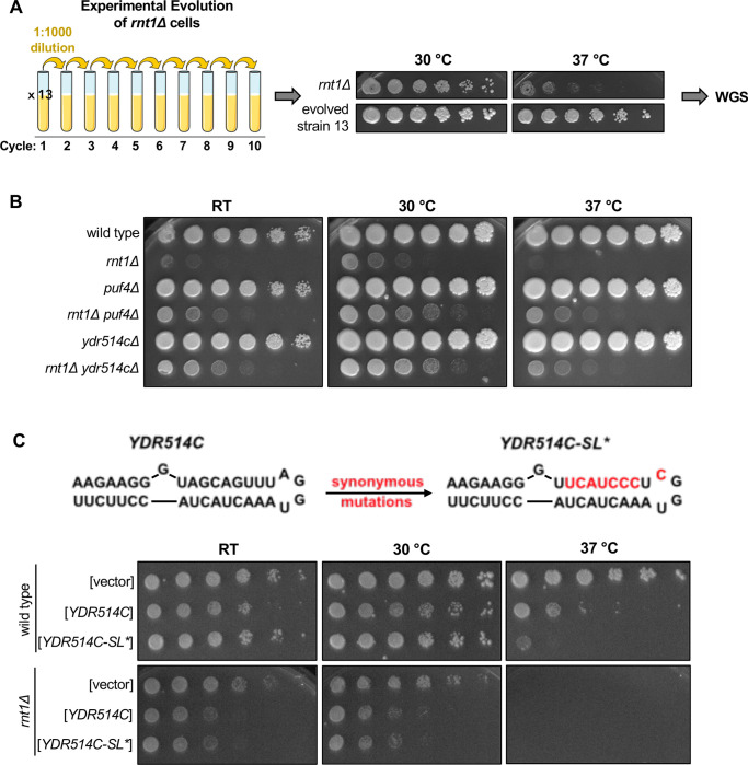
A large fraction of newly transcribed RNA is degraded in the nucleus, but nuclear mRNA degradation pathways remain largely understudied. The yeast nuclear endoribonuclease Rnt1 has a well-characterized role in the maturation of many ncRNA precursors. However, the scope and consequence of its function in mRNA degradation pathways is much less defined. Here, we take a whole-transcriptome approach to identify Rnt1 cleavage sites throughout the yeast transcriptome in vivo, at single-nucleotide resolution. We discover previously unknown Rnt1 cleavage sites in many protein-coding regions and find that the sequences and structures necessary for cleavage mirror those required for the cleavage of known targets. We show that the nuclear localization of Rnt1 functions as an additional layer of target selection control and that cleaved mRNAs are likely exported to the cytoplasm to be degraded by Xrn1. Further, we find that several cleavage products are much more abundant in our degradome sequencing libraries than decapping products, and strikingly, mutations in one Rnt1 target, YDR514C, suppress the growth defect of a RNT1 deletion. Overexpression of YDR514C results in slow growth, further suggesting that Rnt1 may limit the expression of YDR514C to maintain proper cell growth. This study uncovers a broader target range and function for the well-known RNase III enzyme.


Figures

Figure 1:
Figure 1:. PARE identifies known Rnt1 cleavage sites and substrates.
(A) Schematic of PARE workflow. Total RNA is isolated from RNT1 and rnt1Δ strains. T4 RNA ligase ligates an adapter (red rectangle) onto exposed 5’ phosphates (red P’s) resulting from cleavage or decapping. Next-generation sequencing is performed from the 5’ adapter, resulting in reads that begin at the first nucleotide after the cleavage or decapping site. (B) >80,000 sites were detected with reads ≥1 cpm in the RNT1 strain (x-axis). 496 of these were decreased in the rnt1Δ strain (log2(FC)>4 blue, green, and yellow dots). Known ncRNA processing sites are shown in green, and known mRNA sites are shown in yellow. Shown are the averages of two independent biological replicates. (C) 496 putative Rnt1 cleavage sites cluster into 166 different substrates. All known Rnt1 ncRNA targets are detected as well as 2 novel snoRNA targets. 63 mRNA targets are detected, of which 3 are known and 60 are novel. Other sites detected include intergenic, antisense, intronic, and 5’ and 3’ UTR sites. (D) PARE detects known Rnt1 cleavage sites in ncRNA targets, validating PARE as a reliable method for identifying novel Rnt1 cleavage sites. Some known mRNA sites are also detected, but most have reads <1 cpm in RNT1. (E) PARE precisely detects the known Rnt1 cleavage site in pre-SNR83 (red arrowhead) located 61 nts upstream of its mature 5’ end (green arrowhead) on the 5’ side of an AGUU stem loop. PARE additionally reveals a novel site (pink arrowhead) located on the 3’ side of stem. Structure of the stem loop and IGV PARE screenshot are shown. Northern blot using a probe that hybridizes to mature SNR83 (grey bar) was performed in duplicate.
Figure 2:
Figure 2:. PARE identifies novel Rnt1 mRNA targets.
(A-D) PARE screenshots of Rnt1-cleaved mRNA targets. Strong peaks for Rnt1 cleavage (red arrowheads) are detected in (A) the known mRNA target BDF2 and novel targets (B) CAF4, (C) YDR514C, and (D) MTM1. PARE was performed in duplicate and both independent biological replicates are shown. (E) Northern blots detect cleavage products of BDF2 and CAF4. Shown is a representative of two independent biological replicates. PGK1 was used as a loading control. (F) Sequence alignment of 42 of 63 mRNA tetraloops and surrounding sequences.
Figure 3:
Figure 3:. Rnt1 directly and independently cleaves mRNAs.
(A) Validation of Rnt1 catalytic mutant by growth assay and by northern blot of SNR83. Growth assay strains were spotted on SC-Leu. Experiment was performed using two independent biological replicates. (B) Rnt1 catalytic mutant PARE. Wild-type RNT1 or rnt1-D245R cloned into a plasmid, or empty vector, was expressed in a rat1-ts xrn1Δ rnt1Δ triple mutant strain. PARE panels of BDF2, CAF4, and YDR514C are shown. Experiment was performed using three independent biological replicates. (C) Schematic of in vitro PARE workflow: RNA was isolated from a rnt1Δ-only strain and incubated with 0, 4, or 8 pmol of recombinant Rnt1. This RNA was then analyzed by PARE. (D) In vitro Rnt1 cleavage of BDF2, CAF4, and YDR514C (red arrowheads, Rnt1 cleavage sites detected in vivo; grey arrowheads, additional Rnt1 cleavage sites detected in vitro, but not in vivo). In vitro PARE was performed using two independent biological replicates. (E) Comparison of the numbers of in vivo and in vitro targets. (F) In vitro Rnt1 cleavage sites in MIG2 (panels 3–5) compared to in vivo Rnt1 cleavage sites in MIG2 (panels 1–2).
Figure 4:
Figure 4:. Localization is a key determinant in Rnt1 mRNA selection and cleavage.
(A) Confirmation of Rnt1 cytoplasmic relocalization by confocal fluorescence microscopy. RNT1-GFP or rnt1-ΔNLS-GFP cloned into a plasmid, or empty vector, was expressed in a rnt1Δ-only strain. The nucleus was stained with DAPI, pseudo-colored red. Experiment was performed using two independent biological replicates. (B) PARE of BDF2 and CAF4 cleaved by cytoplasmic Rnt1. Wild-type RNT1, rnt1-ΔNLS-GFP, or rnt1-K45I was expressed in a rat1-ts xrn1Δ rnt1Δ triple mutant strain. Experiments were performed using two independent biological replicates. (C) Northern blot of BDF2 and CAF4 cleaved by cytoplasmic Rnt1. PGK1 was used as a loading control.
Figure 5:
Figure 5:. Rnt1-cleaved mRNAs are subsequently degraded by Xrn1.
(A) Rnt1 cleavage peaks in BDF2, CAF4, and YDR514C in xrn1Δ-only PARE data. Two independent biological replicates are shown. PARE dataset from the rat1-ts xrn1Δ double mutant background is also shown for comparison. (B) Northern blot of BDF2 and CAF4 using RNA from wild-type RNT1, rnt1Δ, rat1-ts, and xrn1Δ single mutant strains. Experiment was performed using two independent biological replicates. PGK1 was used as a loading control.
Figure 6:
Figure 6:. Rnt1 and decapping products are derived from mRNAs with distinct poly(A) status.
IGV screenshots of RNT1 vs rnt1Δ PARE data generated from poly(A)-enriched and poly(A)-depleted samples show different distributions for Rnt1 products (red arrowheads) and decapping products (grey bars). The BDF2 mRNA is also a substrate for spliceosome-mediated decay (SMD, purple arrowhead).
Figure 7:
Figure 7:. Rnt1 cleavage of YDR514C mRNA contributes to normal cell growth.
(A) Schematic of rnt1Δ experimental evolution. Thirteen cultures of rnt1Δ were grown to saturation, then sub-cultured to a 1:1000 dilution for 10 cycles. Solid media growth assays were performed, and strains showing enhanced growth compared to the rnt1Δ parent strain were analyzed by whole-genome sequencing (WGS). The growth assay of evolved strain 13 is depicted above. (B) Growth assay confirming enhanced growth of a rnt1Δ ydr514cΔ double mutant compared to rnt1Δ. Experiment was performed using two independent biological replicates. (C) Growth assay confirming impaired growth of wild type and rnt1Δ strains harboring plasmids that overexpress wild-type YDR514C or the ydr514c stem loop mutant. Experiment was performed using two independent biological replicates. Strains were spotted on SC-Leu.

References

    1. Abramson J., Adler J., Dunger J., Evans R., Green T., Pritzel A., Ronneberger O., Willmore L., Ballard A. J., Bambrick J., Bodenstein S. W., Evans D. A., Hung C.-C., O’Neill M., Reiman D., Tunyasuvunakool K., Wu Z., Žemgulytė A., Arvaniti E., … Jumper J. M. (2024). Accurate structure prediction of biomolecular interactions with AlphaFold 3. Nature, 630(8016), 493–500. 10.1038/s41586-024-07487-w - DOI - PMC - PubMed
    1. Allmang C., Kufel J., Chanfreau G., Mitchell P., Petfalski E., & Tollervey D. (1999). Functions of the exosome in rRNA, snoRNA and snRNA synthesis. EMBO Journal, 18(19). 10.1093/emboj/18.19.5399 - DOI - PMC - PubMed
    1. Allmang C., & Tollervey D. (1998). The role of the 3’ external transcribed spacer in yeast Pre-rRNA processing. Journal of Molecular Biology, 278(1). 10.1006/jmbi.1998.1693 - DOI - PubMed
    1. Andrews S. (2010). FastQC: A quality control tool for high throughput sequence data. Http://Www.Bioinformatics.Babraham.Ac.Uk/Projects/Fastqc.
    1. Assenholt J., Mouaikel J., Andersen K. R., Brodersen D. E., Libri D., & Jensen T. H. (2008). Exonucleolysis is required for nuclear mRNA quality control in yeast THO mutants. RNA, 14(11), 2305–2313. 10.1261/rna.1108008 - DOI - PMC - PubMed

Publication types

LinkOut - more resources