[Preprint]. 2025 Aug 14:2025.08.13.25333146.
doi: 10.1101/2025.08.13.25333146.

The TECTB-C225Y Variant Causing Autosomal Dominant Deafness in a Nicaraguan Family Enhances Sensitivity to Noise-Induced Hearing Loss in Mice

Affiliations

The TECTB-C225Y Variant Causing Autosomal Dominant Deafness in a Nicaraguan Family Enhances Sensitivity to Noise-Induced Hearing Loss in Mice

Evan B Hale et al. medRxiv. .

Abstract

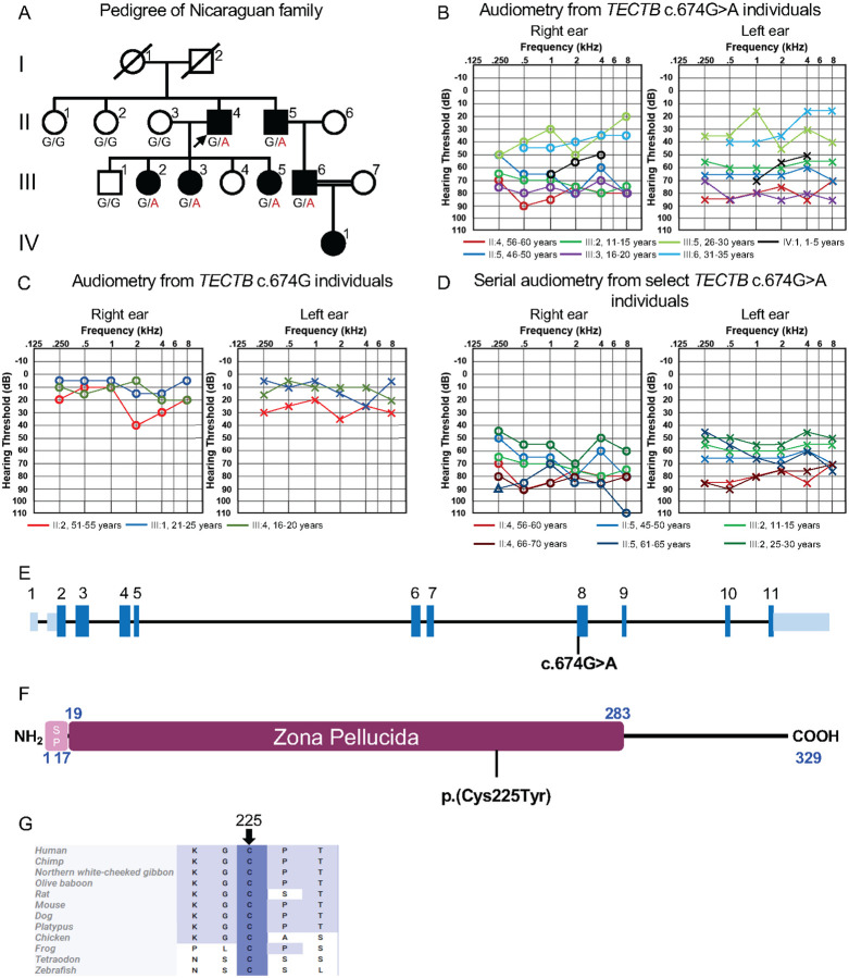
Identifying new genes responsible for non-syndromic hearing loss remains a critical goal, as many individuals with hereditary deafness still lack a molecular diagnosis despite comprehensive genetic testing. The tectorial membrane (TM) is a specialized, collagen-rich, acellular matrix of the inner ear, essential for stimulating mechanosensitive hair cell bundles during sound transduction, and its structural integrity is critical for frequency tuning and auditory sensitivity. Although mutations in genes encoding a number of non-collagenous proteins found in the TM (TECTA, CEACAM16, OTOG, OTOGL) have been identified as deafness genes, definitive evidence implicating β-tectorin (TECTB) in human hearing loss has been lacking. Here, we present multiple lines of genetic and experimental evidence linking a missense variant in TECTB (c.674G>A, p.Cys225Tyr) to autosomal dominant, non-syndromic hearing loss in a multigenerational family. The variant alters one of eight highly conserved cysteines present within the zona pellucida (ZP) domain of TECTB and is predicted to disrupt protein folding and matrix assembly. Using a Tectb-C225Y knock-in mouse model, we show that homozygous animals exhibit severe hearing loss and profound disruption of TM morphology, while heterozygote animals display decreased matrix content within the TM and increased susceptibility to noise-induced hearing loss-despite normal auditory thresholds. These findings identify TECTB as a novel human deafness gene, further elucidate its structural role in maintaining TM integrity, and highlight its contribution to resilience against environmental and age-related auditory decline.

Keywords: Hearing loss; beta-tectorin (TECTB); deafness; striated sheet matrix; tectorial membrane.


Conflict of interest statement

Competing interests The authors declare no competing interests.

Figures

Figure 1.
Figure 1.. TECTB-C225Y Variant Segregates with Non-syndromic Hearing Loss in a Nicaraguan Family.
(A) An abbreviated pedigree of the multigenerational family showing segregation of the heterozygous TECTB c.674G>A, p.Cys225Tyr variant. Filled symbols indicate affected individuals; open symbols indicate unaffected individuals. The proband II:4 is denoted with an arrow. Deceased individuals are indicated by a diagonal line. The variant nucleotide substitution “A” is shown in red. (B) Pure-tone audiograms of affected individuals II:4 (red), II:5 (dark blue), III:2 (dark green), III:3 (purple), III:5 (light green), III:6 (light blue), and IV:1 (black), with corresponding ages at the time of testing in the legend below. Air-conduction thresholds in dB hearing level are represented by circles (right ear) and crosses (left ear). (C) Pure-tone audiograms of unaffected individuals II:2 (red), III:1 (blue), and III:4 (green) carrying the reference allele, with ages at the time of evaluation presented in the legend below. Symbols as in panel B. (D) Follow-up pure-tone audiograms from II:4, II:5 and III:2 that span 12, 16, and 16 years, respectively. (E) Schematic representation of the TECTB (NM_058222.3), with coding exons shown in dark blue boxes and untranslated sequences in light blue. The identified variant maps to exon 8. (F) Domain structure of the TECTB protein, showing the signal peptide (SP, light maroon), and the conserved zona pellucida domain (ZP, maroon). The location of the C225Y substitution is indicated with a black bar. (G) Multiple sequence alignment of TECTB orthologues highlighting the conserved cysteine residue at position 225 (arrow), substituted in the affected family.
Figure 2.
Figure 2.. TectbC225Y/C225Y Mice Exhibit a Severe Hearing Deficit.
(A) Auditory brainstem response (ABR) and (B) distortion product otoacoustic emission (DPOAE) thresholds of Tectb-C225Y mice tested at 5 weeks, and 3, 6, 9, 12, and 18 months of age. Data are shown as individual values with an overlaid mean ± SEM. Upward-pointing arrows indicate that at the highest sound pressure level tested (80 dB SPL), at least one mouse in the group had no detectable response at that frequency; these values were entered as 85 dB for analysis. TectbC225Y/C225Y mice exhibited significantly higher ABR and DPOAE thresholds than both TectbC225Y/+ (blue asterisks) and wild type (black asterisks) mice across all tested ages. No significant differences were observed between TectbC225Y/+ and wild-type mice at all but one data point (purple asterisk). The number of animals per group is indicated within each plot. Statistical analysis: two-way ANOVA with a matched design between frequencies for each mouse. Šidák’s multiple comparison test was to evaluate differences between genotypes at each frequency; ns – not significant, * p<0.05, ** p<0.01, *** p<0.001, **** p<0.0001.
Figure 3.
Figure 3.. TM morphology is disrupted in mice expressing TECTB-C225Y.
(A) Toluidine blue-stained 1 μm resin sections from the 8 and 20 kHz cochlear regions of wild-type, TectbC225Y/+ and TectbC225Y/C225Y mice at 2–3 months of age. (B) Representative images showing the adaptive thresholding method used to quantify TM cross-sectional area and staining density. Images correspond to the 8 kHz region from 2–3-month-old mice shown in A. Cyan, unstained portion of TM. Magenta, stained portion of TM. (C) Example of a cochlear cross-section from the 4 kHz region of a 14-month-old TectbC225Y/C225Y mouse, illustrating fusion of the disrupted TM with Reissner’s membrane (red arrow). Scale bars: 100 μm. (D) Quantification of TM cross-sectional area across genotypes and regions. (E) Percentage of the TM cross-sectional area that is unstained. Values for TectbC225Y/C225Y TMs at 4 kHz (panels D-E), indicated in pink, were aberrated by attachment of the TM to Reissner’s membrane. The inclusion of these values did not affect the statistical significance levels calculated. Statistical analysis: One-way ANOVA was performed for each frequency, followed by Šidák’s multiple comparisons test between genotypes with Benjamini-Hochberg correction applied within each age group. Percentage of unstained area values were arcsine square root-transformed prior to statistical analysis. ns – not significant, * p<0.05, ** p<0.01, *** p<0.001.
Figure 4.
Figure 4.. TECTB-C225Y disrupts TM ultrastructure.
(A) Representative micrographs of toluidine blue-stained 1 μm resin sections from the 40 kHz region of wild-type, TectbC225Y/+ and TectbC225Y/C225Y cochleae. Arrowheads indicate Hensen’s stripe, arrows indicate the marginal band. (B) and (C), pooled morphology scores from blinded reviewers assessing the integrity of the marginal band (B) and Hensen’s stripe (C) in TM cross-sections from the 40 kHz region. Scoring was based on structural continuity and deviation from typical shape. The number of animals per group is indicated within each plot.
Figure 5.
Figure 5.. TectbC225Y/C225Y tectorial membranes lack striated sheet matrix.
Transmission electron micrographs from the central body of tectorial membranes in the 4 kHz region of 2–3-month-old mice. Arrows point to collagen fibers; arrowheads point to striated sheet matrix. Scale bars, 400 nm (main panels) and 100 nm (insets).
Figure 6:
Figure 6:. TectbC225Y/+ Mice show increased vulnerability to noise damage.
Threshold shifts in ABR and DPOAE measurements at 16 weeks (A) and 18 months (B) of age following noise exposure. Threshold shifts were calculated by subtracting pre-exposure values from those obtained 2 days and 14 days post-exposure. Wild-type and TectbC225Y/+ mice were exposed for 2 hours to 8–16 kHz octave-band noise at 100 dB SPL, indicated by the shaded area on each graph. When no response was detected at 80 dB SPL, the threshold was entered as 85 dB, one interval above the system’s test ceiling. Data are shown as mean ± SEM. Statistical Analysis: two-way ANOVA followed by Šidák’s multiple comparisons test between genotypes at each frequency. *p < 0.05, **p < 0.01, ***p < 0.001, ****p < 0.0001.

References

    1. Adler H. J., Niemiec A. J., Moody D. B., & Raphael Y. (1995). Tectorial membrane regeneration in acoustically damaged birds: an immunocytochemical technique. Hear Res, 86(1–2), 43–46. 10.1016/0378-5955(95)00051-5 - DOI - PubMed
    1. All of Us Research Program Genomics, I. (2024). Genomic data in the All of Us Research Program. Nature, 627(8003), 340–346. 10.1038/s41586-023-06957-x - DOI - PMC - PubMed
    1. Armstrong N. M., Wang H., E J. Y., Lin F. R., Abraham A. G., Ramulu P., Resnick S. M., Tian Q., Simonsick E., Gross A. L., Schrack J. A., Ferrucci L., & Agrawal Y. (2022). Patterns of Prevalence of Multiple Sensory Impairments Among Community-dwelling Older Adults. J Gerontol A Biol Sci Med Sci, 77(10), 2123–2132. 10.1093/gerona/glab294 - DOI - PMC - PubMed
    1. Azaiez H., Decker A. R., Booth K. T., Simpson A. C., Shearer A. E., Huygen P. L., Bu F., Hildebrand M. S., Ranum P. T., Shibata S. B., Turner A., Zhang Y., Kimberling W. J., Cornell R. A., & Smith R. J. (2015). HOMER2, a stereociliary scaffolding protein, is essential for normal hearing in humans and mice. PLoS Genet, 11(3), e1005137. 10.1371/journal.pgen.1005137 - DOI - PMC - PubMed
    1. Bokhove M., & Jovine L. (2018). Structure of Zona Pellucida Module Proteins. Curr Top Dev Biol, 130, 413–442. 10.1016/bs.ctdb.2018.02.007 - DOI - PubMed

Publication types

LinkOut - more resources