. 2025 Aug 26;122(34):e2507221122.
doi: 10.1073/pnas.2507221122. Epub 2025 Aug 20.

A β-cap on the FliPQR protein-export channel acts as the cap for initial flagellar rod assembly

Affiliations

A β-cap on the FliPQR protein-export channel acts as the cap for initial flagellar rod assembly

Miki Kinoshita et al. Proc Natl Acad Sci U S A. .

Abstract

The FliPQR complex constitutes a channel for export of the flagellar proteins involved in axial structure assembly. It also serves as a template for the assembly of the rod structure, which consists of FliE, FlgB, FlgC, FlgF, and FlgG. FliP, FliQ, and FliR assemble into a right-handed helical structure within the central pore of the flagellar basal body MS-ring, and the complex has two gates on the cytoplasmic and periplasmic sides. The periplasmic gate, formed by the N-terminal α-helices of FliP and FliR, remains closed until six FliE subunits assemble onto FliP and FliR to form the first layer of the rod, but it has remained unclear how each FliE subunit opens the gate and assembles in the absence of the rod cap required for efficient assembly of other rod proteins. Here, we present a cryoelectron microscopy structure of the FliPQR complex in closed form at 3.0 Å resolution. A β-cap, formed by the N-terminal β-strands of FliP and FliR, is located at the top of the FliPQR complex and tightly seals the closed gate. The β-cap has a narrow pore that efficiently and accurately leads the first FliE subunit to its assembly site. Interactions of FliE with FliP and FliR induce a conformational change in FliP and FliR, with their N-terminal α-helices move up and outward to open the gate. Consequently, each of the N-terminal β-strands of FliP and FliR detaches from the β-cap one after another, thereby creating a docking site for the next FliE subunit to efficiently assemble.

Keywords: cryoelectron microscopy; flagellar rod; flagellum; type III secretion system.


Conflict of interest statement

Competing interests statement:The authors declare no competing interest.

Figures
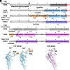
Fig. 1.
Fig. 1.
Amino acid sequences and atomic models of the N-terminal regions of FliP and FliR in the completely closed form of the FliPQR complex. (A and B) Amino acid sequences of the N-terminal regions of (A) FliP and (B) FliR. The N-terminal regions of FliP and FliR are in the closed conformation in the 6R69 and 9K29 structures, but in the open conformation in the 8WKK structure. The first 21 amino acid residues of FliP serve as the N-terminal signal peptide that is cleaved during membrane insertion. The arrow indicates that the cleavage occurs between Ala-21 and Gln-22. Residues 22 to 42 in the 6R69 structure and residues 22 to 35 in the 8WKK structure are invisible. However, these residues are visible in the 9K29 structure. The well-conserved MSTF motif is highlighted in red. The first 5 and 3 residues of FliR are not visible in the 6R69 and 8WKK structures, respectively, but are visible in the 9K29 structure. These visible regions, highlighted in orange, form the β-cap, which is stabilized by six tryptophan residues (Trp-38 of FliP and Trp-9 of FliR) indicated by open boxes. (C) Cα ribbon diagrams of the atomic models of two FliP subunits, the first (FliP1) and second (FliP2), obtained in this study (PDB ID: 9K29). (D) Cα ribbon diagram of the atomic model of FliR obtained in this study (PDB ID: 9K29).
Fig. 2.
Fig. 2.
The cryoEM structure of the FliPQR complex reconstituted in the peptidisc. (A) Cα ribbon diagram of the atomic model of the FliPQR complex with a stoichiometry of 5 FliP, 4 FliQ, and 1 FliR subunits (PDB ID: 9K29). FliP, FliQ, and FliR are colored in sky blue, dark sea green, and plum, respectively. The extreme N-terminal regions of FliP and FliR, which are colored in orange, form the β-cap. The p-loop, consisting of residues 155 to 166, is located in the outermost part of the FliPQR complex. (B) The β-cap formed by the N-terminal β strands of FliP and FliR. The FliP subunit located at the top of the right-handed helical structure of the FliPQR complex is designated as the first FliP (FliP1) subunit. The remaining subunits, arranged along the helical staircase, are subsequently referred to as the second (FliP2), third (FliP3), fourth (FliP4), and fifth (FliP5) subunits, respectively. Four FliP subunits, FliP2, FliP3, FliP4, and FliP5, but not FliP1, contain two β-strands (β1, β2) forming a β-hairpin at their N-terminal regions. FliR possesses a β-strand (β1) in its extreme N-terminal region. The four β-hairpins and β1 of FliR form the β-cap, which is stabilized by hydrophobic interactions of six tryptophan residues (Trp-38 of FliP and Trp-9 of FliR). The extreme N-terminal region of α1 of FliP1 project into the cavity of the β-cap. (C) The narrow pore within the β-cap. The N-terminal region of FliP1 is not involved in the β-hairpin formation, which leaves a narrow pore between the FliR and FliP5 subunits (Left). Comparison of the 9K29 structure with the 8WKK structure by superposition reveals that the pore is sufficiently wide to accommodate helix α3 of the first FliE subunit (FliE1, yellow), which is inserted between FliR and FliP5 in the 8WKK structure (Right). Side-chain atoms of FliP and FliR involved in the formation of the β-cap are indicated by orange spheres. The R-loop formed by residues 55 to 68 of FliR extends into the pore, where it exhibits an apparent steric clash with the α-helix as marked by a red circle.
Fig. 3.
Fig. 3.
Structural comparisons of FliP and FliR subunits in the FliPQR complex of the present study (PDB ID: 9K29) with those of previous studies. (A) Structural comparisons of the five FliP subunits in the three different FliPQR structures. The RMS deviations (RMSDs) for these superpositions are provided in SI Appendix, Table S2. The conformations of the five FliP subunits are slightly different from each other in the 6R69 (dark gray) and 9K29 (sky blue) structures, whereas the five FliP subunits are nearly identical to each other in the 8WKK structure (royal blue). In the 9K29 structure, the N-terminal β-hairpin structure is visible in FliP2, FliP3, FliP4, and FliP5, but not in FliP1. The orientation of α1 is different among the five FliP molecules in the 6R69 and 9K29 structures. In the FliP5 subunit, the helix–turn–helix structure (HTHα3-α4) formed by α3 and α4 and a loop connecting these two helices is shifted outward relative to the other four FliP subunits. In the 8WKK structure, the orientation of α1 and the position of HTHα3-α4 are nearly identical among the five FliP molecules. (B) Structural comparison of the FliR subunits in the three different FliPQR structures. The FliR subunits from the 6R69 and 8WKK structures were individually superimposed onto the equivalent coordinate in the 9K29 structure. The RMSD values for these superpositions are listed in SI Appendix, Table S3. The overall structure of FliR is almost identical among the 6R69 (silver), 9K29 (plum), and 8WKK (purple) structures. However, in the 8WKK structure, the N-terminal α1 helix adopts a different orientation compared to those in the 6R69 and 9K29 structures. The N-terminal β-strand (β1) is visible only in the 9K29 structure.
Fig. 4.
Fig. 4.
Mutational analysis of the conserved MTSF motif in FliP. (A) Evolutionarily conserved residues of FliP. Conservation scores were calculated using the ConSurf web server. Residues are colored according to sequence conservation among 150 bacterial species. (B) Motility assay of a Salmonella fliP null mutant carrying pTrc99AFF4 (∆fliP), pKY69 (WT), pMKM69(∆MTSF) (∆MTSF), pMKM69(T62A/S63A) (T62A/S63A), or pMKM69(T62G/S63G) (T62G/S63G) in soft agar. The plates were incubated at 30 °C for 6 h. At least seven independent assays were performed. (C) Secretion assay. Whole-cell proteins (Cell) and culture supernatants (Sup) were prepared from the above strains. A 5 μL aliquot of each protein sample, normalized to OD600, was subjected to SDS-PAGE, followed by immunoblotting with polyclonal anti-FlgD (first row) and anti-FliP (second row) antibodies. Molecular mass markers (kDa) are shown on the Left. At least three independent assays were carried out.
Fig. 5.
Fig. 5.
Role of the conserved Leu-92 residue of FliP in flagellar protein export. (A) Evolutionarily conserved residues of FliP. Conservation scores were calculated using the ConSurf web server (https://consurf.tau.ac.il/consurf_index.php). Residues are colored according to sequence conservation among 150 bacterial species. (B) Location of Leu-92 in the hydrophobic side-chain interaction networks in the closed (Left) and open (Right) conformations of the FliPQR complex. The interaction between FliE and FliP causes a conformational transition from the closed to the open state of FliP. Leu-92 forms an intramolecular hydrophobic interaction network with Phe-71, Leu-96, Met-236, and Leu-239. The FliE–FliP interaction induces remodeling of the hydrophobic interaction network formed by Phe-71, Leu-92, Leu-96, Met-236, and Leu-239 (Upper). Leu-92 of FliP1 also makes intermolecular hydrophobic contacts with both Pro-172 and Val-175 of FliP2 in the open form, but only with Val-175 in the closed form (Lower). (C) Motility assay of a Salmonella fliP null mutant carrying pTrc99AFF4 (indicated as ∆fliP), pKY69 (indicated as WT), or pMKM69(L92A) (indicated as L92A) in soft agar. The plates were incubated at 30 °C. At least seven independent assays were carried out. (D) Secretion assay. Whole-cell proteins (Cell) and culture supernatants (Sup) were prepared from the above strains. A 5 μL aliquot of each protein sample, normalized to OD600, was subjected to SDS-PAGE, followed by immunoblotting with polyclonal anti-FlgD (first row) and anti-FliP (second row) antibodies. Molecular mass markers (kDa) are shown on the Left. At least three independent experiments were performed. (E) Location of intragenic suppressor mutation sites identified in up-motile mutants isolated from the fliP(L92A) mutant. The L92A mutation site and its intragenic suppressor mutation sites are highlighted in red and blue, respectively. Pro-30 is located within the β-cap in the closed structure. Leu-45 forms a hydrophobic core with surrounding residues in helix α1, contributing to the closure of the periplasmic gate. Leu-90 engages in hydrophobic interaction with Phe-64 in the MSTF motif when FliP is in the closed form. Leu-45 and Leu-90 are also involved in the FliP–FliE interaction in the open structure (Also see SI Appendix, Fig. S6B). Arg-168 of FliP1 forms a hydrogen bond with the carbonyl oxygen of Phe-99 of FliP2 in the closed conformation but not in the open conformation.
Fig. 6.
Fig. 6.
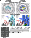
Effect of in-frame deletions of the p-loop of FliP on flagellar protein export. (A) Structural comparison of the p-loop conformations in the closed (PDB ID: 9K29) and open (PDB ID: 8WKK) forms of the FliPQR complex. The open and closed structures are superimposed on the equivalent coordinates of the 7NVG structure. FliP, FliQ, and FliR are colored in sky blue, dark sea green, and plum, respectively, in the 9K29 structure. FliE, FliP, FliQ, and FliR are colored in yellow, royal blue, green, and purple, respectively, in the 8WKK structure. The FliPQR complex is located within the central pore of the MS-ring. Each p-loop (residues 155 to 166) makes physical contact with the i-loop of the MS-ring protein FliF (red, residues 159 to 172) within the MS-ring after 6 FliE subunits assemble on FliP and FliR. (B) Motility assay of a Salmonella fliP null mutant carrying pTrc99AFF4 (indicated as ∆fliP), pKY69 (indicated as WT), pMKM69(∆155-164) (indicated as ∆155-164), pMKM69(∆155-165) (indicated as ∆155-165), pMKM69(∆156-163) (indicated as ∆156-163), or pMKM69(∆157-162) (indicated as ∆157-162) in soft agar. The plates were incubated at 30 °C for 7 h. At least seven independent assays were carried out. (C) Secretion assay. Whole-cell proteins (Cell) and culture supernatants (Sup) were prepared from the above transformants. A 5 μL aliquot of each protein sample, normalized to OD600, was subjected to SDS-PAGE, followed by immunoblotting with polyclonal anti-FlgD (first row) and anti-FliP (second row) antibodies. Molecular mass markers (kDa) are shown on the Left. At least three independent assays were performed.
Fig. 7.
Fig. 7.
FliE assembly mechanism. The periplasmic gate of the FliPQR complex is completely closed by the β-cap while leaving a narrow pore (I), as shown in the CPK representation. The first FliE subunit (E1) is transported into the export channel by the fT3SS, and α3 of FliE1 inserts into the narrow pore of the β-cap. The interaction between the closed form of FliR (RC) and FliE1 induces a conformational change of α1 of FliR, allowing this helix not only to adopt an open conformation (RO) but also to form the D0-like domain together with α3 of FliE1 (SI Appendix, Fig. S14). Then, α3 of FliE1 associates with the closed form of the FliP5 subunit (P5C), thereby inducing a conformational change of the conserved MSTF motif of FliP (SI Appendix, Fig. S15). As a result, FliP5 adopts an open conformation (P5O), creating the next insertion site (II) for the second FliE subunit (FliE2) within the β-cap. This open conformation is stabilized by the direct contact of the p-loop of FliP5O with the inner wall of the MS-ring. When FliE2 inserts into this site, helices α2 and α3 of FliE2 form domain D0 between FliP5O and the closed form of FliP4 (P4C). The FliE2–FliP4C interaction allows FliP4C to adopt the open conformation (P4O). The FliE assembly proceeds by repeating these steps. When the sixth FliE subunit (FliE6) inserts between FliP1O and FliRO, the periplasmic gate of the FliPQR complex is completely opened, allowing other flagellar structural subunits to go through its channel, diffuse through the central channel of the growing flagellum, and assemble at the distal end (Movie S2).

Similar articles

References

    1. Minamino T., Kinoshita M., Structure, assembly, and function of flagella responsible for bacterial locomotion. EcoSal Plus 11, eesp00112023 (2023). - PMC - PubMed
    1. Minamino T., Kinoshita M., Morimoto Y. V., Namba K., Activation mechanism of the bacterial flagellar dual-fuel protein export engine. Biophys. Physicobiol. 19, e190046 (2022). - PMC - PubMed
    1. Minamino T., Kinoshita M., Namba K., Insight into distinct functional roles of the flagellar ATPase complex for flagellar assembly in Salmonella. Front. Microbiol. 13, 864178 (2022). - PMC - PubMed
    1. Minamino T., Namba K., Distinct roles of the FliI ATPase and proton motive force in bacterial flagellar protein export. Nature 451, 485–488 (2008). - PubMed
    1. Paul K., Erhardt M., Hirano T., Blair D. F., Hughes K. T., Energy source of flagellar type III secretion. Nature 451, 489–492 (2008). - PubMed

MeSH terms

Substances

LinkOut - more resources