. 2025 Aug 20;20(8):e0330229.
doi: 10.1371/journal.pone.0330229. eCollection 2025.

The role of cyclic di-GMP in biomaterial-associated infections caused by commensal Escherichia coli

Affiliations

The role of cyclic di-GMP in biomaterial-associated infections caused by commensal Escherichia coli

Shanshan Li et al. PLoS One. .

Abstract

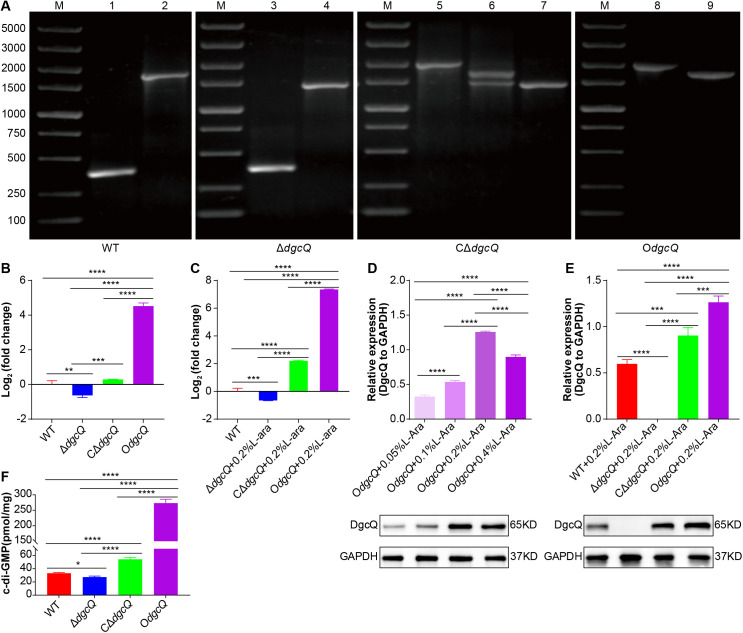
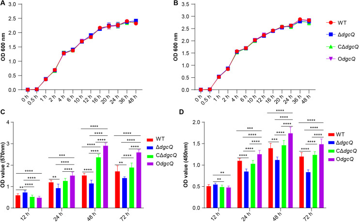
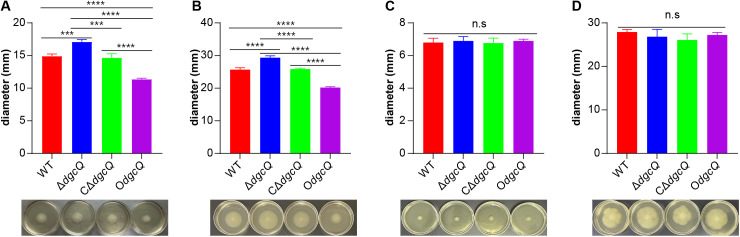
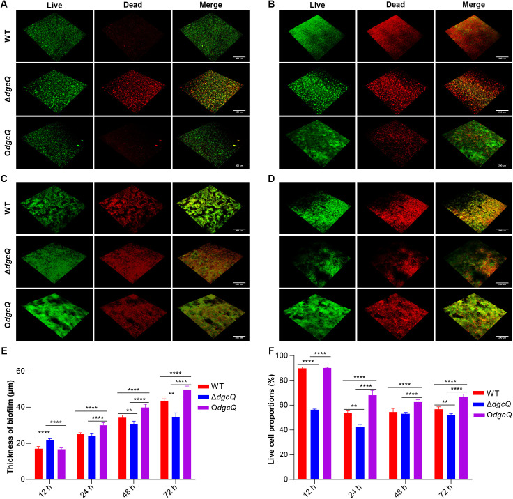
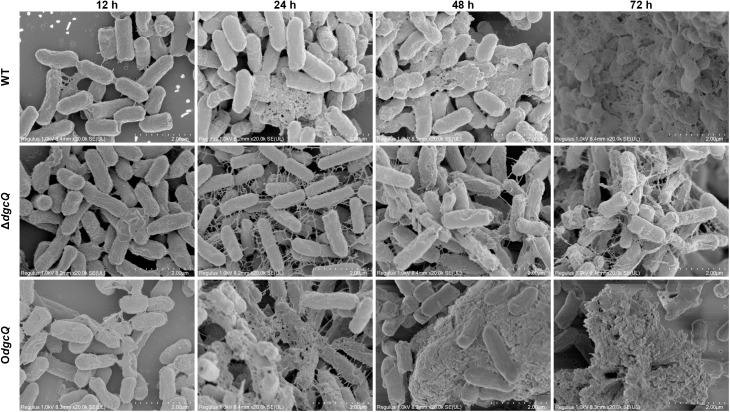
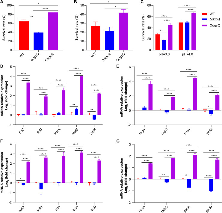
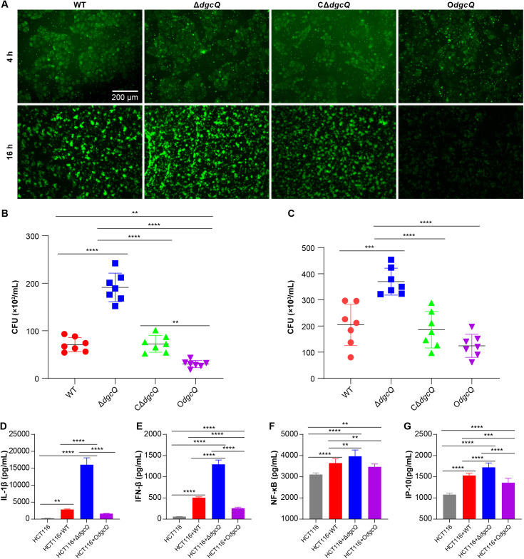
Biofilms are protective structures that bacteria use to evade the immune system and resist antibiotics, leading to complications in medical treatments, especially with implanted devices. The molecule cyclic di-GMP (c-di-GMP) is crucial for biofilm formation in Escherichia coli (E. coli). To understand its role in biomaterial-associated infections (BAIs), we created four E. coli strains with varying c-di-GMP levels: a knockout strain (ΔdgcQ), an overexpression strain (OdgcQ), a complemented strain (CΔdgcQ), and a wild-type mutant strain (WT). By employing in vitro BAI models and techniques such as crystal violet (CV) staining, XTT assay, confocal laser scanning microscopy (CLSM), and scanning electron microscopy (SEM), we observed that the ΔdgcQ strain, with low c-di-GMP levels, adhered more readily to biomaterial surfaces at the initial stage of biofilm formation, yet faced difficulties in sustaining mature biofilms. In contrast, OdgcQ and CΔdgcQ with higher c-di-GMP were able to generate more mature biofilms on biomaterial surfaces. Additionally, c-di-GMP was found to negatively regulate bacterial swimming motility and enhance the ability to cope with environmental stresses. The results also reiterate the canonical function of c-di-GMP, which is to reduce the motility of bacteria. Concurrently, gene expression analysis confirmed these findings, revealing that genes related to motility (flhC, flhD, motA, motB, ycgR), extracellular polymeric substances (EPS) synthesis (csgA, csgD, bcsA, ynfM), and stress resistance (sodA, katE, rstA, ibpA, ibpB, hdeA, hdeD, gadA, gadB) were consistently up-regulated in OdgcQ with high c-di-GMP levels. Importantly, ΔdgcQ considerably promoted the adhesion to and invasion of host cells and elicited a stronger host immune response, whereas OdgcQ impaired the ability to interact with host cells, as evidenced by decreased adhesion/invasion and inhibited release of inflammatory cytokines (IL-1β, IFN-β, IP-10, and NF-κB). Collectively, our findings shed light on the c-di-GMP signaling pathway's role in BAIs and propose that modulating this pathway could be a promising strategy for combating E. coli-induced BAIs.


Conflict of interest statement

The authors have declared that no competing interests exist.

Figures

Fig 1
Fig 1. Construction and identification of WT, ΔdgcQ, CdgcQ, and OdgcQ strains.
(A) PCR identification of the WT, ΔdgcQ, CΔdgcQ, and OdgcQ strains. Lane1, 3, 5, 8 amplified by primers P7/8 (expected sizes: 378 bp, 378 bp, 1974 bp, 1974 bp). Lane2, 4, 6, 9 amplified by primers P1/4 (expected sizes: 1695 bp, 1425 bp, 1695/1425 bp, 1695 bp). Lane7 amplified by primers P1/9 (expected sizes: 1445 bp). M: 5000 bp ladder. (B) qRT-PCR identification of the dgcQ mRNA of WT, ΔdgcQ, CΔdgcQ, and OdgcQ strains (n = 3). (C) qRT-PCR identification of the dgcQ mRNA of WT, ΔdgcQ, CΔdgcQ, and OdgcQ strains induced by 0.2% L-arabinose (n = 3). (D) The DgcQ expression of OdgcQ strain induced by various concentrations of L-arabinose (n = 3). (E) The DgcQ expression of WT, ΔdgcQ, CΔdgcQ, and OdgcQ strains induced by 0.2% L-arabinose (n = 3). (F) The c-di-GMP levels of WT, ΔdgcQ, CΔdgcQ, and OdgcQ strains (n = 3). Statistical analysis was performed by One-way ANOVA with LSD (B, C, D, and E) or Tamhane’s T2 (F) multiple comparison test. The values obtained in the WT strain were used as control in qRT-PCR assays (B, C). The DgcQ protein levels were shown relative to GAPDH (D, E). *P< 0.05; **P< 0.01; ***P< 0.001; ****P< 0.0001. Error bars indicated standard deviations.
Fig 2
Fig 2. Biological characteristics analysis of the four mutant strains.
(A) Growth curve of WT, ΔdgcQ, CΔdgcQ, and OdgcQ strains as determined by optical density (OD 600 nm) (n = 3). (B) Growth curve of WT, ΔdgcQ, CΔdgcQ, and OdgcQ strains induced by 0.2% L-arabinose as determined by optical density (OD 600 nm) (n = 3). (C) The biofilm biomass of WT, ΔdgcQ, CΔdgcQ, and OdgcQ strains on PVC (n = 3). (D) The bacterial viability of WT, ΔdgcQ, CΔdgcQ, and OdgcQ biofilms on PVC (n = 3). Statistical analysis was performed by One-way ANOVA with Tukey’s HSD multiple comparison test for all the assays. ** P< 0.01; *** P< 0.001; **** P< 0.0001. Error bars indicated standard deviations.
Fig 3
Fig 3. Comparison of the swimming and swarming ability among the four mutant strains.
(A) Swimming ability at 6 h (n = 3). (B) Swimming ability at 12 h (n = 3). (C) Swarming ability at 12 h (n = 3). (D) Swarming ability at 24 h (n = 3). Statistical analysis was performed by One-way ANOVA with LSD multiple comparison test for all the assays. ***P< 0.001; ****P< 0.0001. n.s, no significance. Error bars indicated standard deviations.
Fig 4
Fig 4. Thickness and live cell proportions of biofilms on PVC observed by CLSM.
(A) Observation at 12 h. (B) Observation at 24 h. (C) Observation at 48 h. (D) Observation at 72 h. (E) The thickness of biofilms at 12, 24, 48, 72 h (n = 5). (F) The live cell proportions of biofilms at 12, 24, 48, 72 h (n = 5). Green represented live cell, red represented dead cell. Scale bar: 200 μm. Statistical analysis was performed by One-way ANOVA with LSD multiple comparison test for all the assays. **P< 0.01; ****P< 0.0001. Error bars indicated standard deviations.
Fig 5
Fig 5. Microstructure of biofilms on PVC observed by SEM at 12, 24, 48, and 72 h.
Scale bar: 2.0 μm.
Fig 6
Fig 6. The adaptation to environmental stresses and related gene expression.
(A) The adaptation to oxidative stress (n = 3). (B) The adaptation to heat stress (n = 3). (C) The adaptation to acid stress (n = 3). (D) Expression of genes related to motility (n = 3). (E) Expression of genes related to EPS (n = 3). (F) Expression of genes related to oxidative and heat stress (n = 3). (G) Expression of genes related to acid stress (n = 3). Statistical analysis was performed by One-way ANOVA with LSD multiple comparison test for all the assays. *P< 0.05; **P< 0.01; ***P< 0.001; ****P< 0.0001. The values obtained in the WT strain were used as control in qRT-PCR assays (D-G). Error bars indicated standard deviations.
Fig 7
Fig 7. The adherence and invasion of the mutant strains to HCT116 and inflammatory response.
(A) Observation of the co-culture system of the four mutant strains and HCT116 cells by fluorescence microscope. Green represented E. coli. Scale bar: 200 μm. (B) The adherence of E. coli to HCT116 (n = 7). (C) The invasion of E. coli to HCT116 (n = 7). (D) ELISA of IL-1β (n = 3). (E) ELISA of IFN-β (n = 3). (F) ELISA of NF-κb (n = 3). (G) ELISA of IP-10 (n = 3). Statistical analysis was performed by One-way ANOVA with LSD (D, E, and G) or Tamhane’s T2 (F) multiple comparison test. **P< 0.01; ***P< 0.001; ****P< 0.0001. Error bars indicated standard deviations.

Similar articles

References

    1. Mori T, Yamamoto H, Tabata T, Shimizu T, Endo Y, Hanasawa K, et al. A free radical scavenger, edaravone (MCI-186), diminishes intestinal neutrophil lipid peroxidation and bacterial translocation in a rat hemorrhagic shock model. Crit Care Med. 2005;33(5):1064–9. doi: 10.1097/01.ccm.0000162952.14590.ec - DOI - PubMed
    1. Earley ZM, Akhtar S, Green SJ, Naqib A, Khan O, Cannon AR, et al. Burn Injury Alters the Intestinal Microbiome and Increases Gut Permeability and Bacterial Translocation. PLoS One. 2015;10(7):e0129996. doi: 10.1371/journal.pone.0129996 - DOI - PMC - PubMed
    1. Ponziani FR, Zocco MA, Cerrito L, Gasbarrini A, Pompili M. Bacterial translocation in patients with liver cirrhosis: physiology, clinical consequences, and practical implications. Expert Rev Gastroenterol Hepatol. 2018;12(7):641–56. doi: 10.1080/17474124.2018.1481747 - DOI - PubMed
    1. Hodges NA, Sussman EM, Stegemann JP. Aseptic and septic prosthetic joint loosening: Impact of biomaterial wear on immune cell function, inflammation, and infection. Biomaterials. 2021;278:121127. doi: 10.1016/j.biomaterials.2021.121127 - DOI - PubMed
    1. Buetti N, Souweine B, Mermel L, Mimoz O, Ruckly S, Loiodice A, et al. Obesity and risk of catheter-related infections in the ICU. A post hoc analysis of four large randomized controlled trials. Intensive Care Med. 2021;47(4):435–43. doi: 10.1007/s00134-020-06336-4 - DOI - PubMed

MeSH terms

LinkOut - more resources