Determination of miRNA in tear extracellular vesicles significantly associated with treatment-requiring retinopathy of prematurity: a pilot study
- PMID: 40835875
- PMCID: PMC12368132
- DOI: 10.1038/s41598-025-15856-2
Determination of miRNA in tear extracellular vesicles significantly associated with treatment-requiring retinopathy of prematurity: a pilot study
Abstract
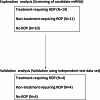
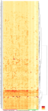
Retinopathy of prematurity (ROP) develops in some premature infants and may be characterized by permanent severe retinal damage necessitating early detection and prompt treatment. The purpose of this study was to investigate whether specific miRNAs in tear extracellular vesicles (EVs) are associated with the development of ROP requiring treatment. Tear samples were collected from 47 infants, including 33 with ROP and 14 without ROP; of the ROP group, 18 infants required treatment. The miRNAs expressed in EVs were profiled by real-time PCR array. An exploratory analysis of differential miRNA expression using tear EV samples from 35 infants was performed. Network analysis conducted for the miRNAs for ROP requiring treatment revealed critical networks of miRNAs linked to IGF1R and VEGF. A machine learning study utilizing the random forest model identified 13 miRNAs with high importance score for the treatment-requiring ROP eyes. After adjustments for birth weight, miR-520a-5p was identified as a candidate marker. Network analysis confirmed a significant association of miR-520a-5p with the VEGF-centered network. When the Gradient boosting decision tree was applied, miR-520a-5p discriminated treatment-requiring ROP with accuracy of 77.8% and an area under the curve (AUC) of 0.889. For the validation phase, 12 unanalyzed infants were examined for the diagnostic accuracy of miR-520a-5p, and the findings confirmed a high diagnostic accuracy of 91.7% and AUC of 0.875 for treatment-required ROP eyes. Notably, miR-520a-5p expression was strongly influenced by infant immaturity, as reflected by gestational age. These findings provide new insights into ROP pathophysiology and suggest that tear-derived miRNAs, particularly those in EVs, may serve as potential biomarkers and inform future therapeutic strategies.
Keywords: Exosome; Extracellular vesicle; Retinopathy of prematurity; Tear; miRNA.
© 2025. The Author(s).
Conflict of interest statement
Declarations. Competing interests: This work was supported by JSPS KAKENHI Grant Number 24K12783 and 23K15931 (T.B. and R.U.). All the remaining authors declare no conflict of interest.
Figures


Similar articles
-
Alteration of Tear Metabolomics Profiling in Infants With Retinopathy of Prematurity.Invest Ophthalmol Vis Sci. 2025 Jun 2;66(6):61. doi: 10.1167/iovs.66.6.61. Invest Ophthalmol Vis Sci. 2025. PMID: 40540260 Free PMC article.
-
Tear Proteomics in Infants at Risk of Retinopathy of Prematurity: A Feasibility Study.Transl Vis Sci Technol. 2024 May 1;13(5):1. doi: 10.1167/tvst.13.5.1. Transl Vis Sci Technol. 2024. PMID: 38691083 Free PMC article.
-
Anti-vascular endothelial growth factor (VEGF) drugs for treatment of retinopathy of prematurity.Cochrane Database Syst Rev. 2018 Jan 8;1(1):CD009734. doi: 10.1002/14651858.CD009734.pub3. Cochrane Database Syst Rev. 2018. PMID: 29308602 Free PMC article.
-
Anti-vascular endothelial growth factor (VEGF) drugs for treatment of retinopathy of prematurity.Cochrane Database Syst Rev. 2016;2:CD009734. doi: 10.1002/14651858.CD009734.pub2. Epub 2016 Feb 27. Cochrane Database Syst Rev. 2016. Update in: Cochrane Database Syst Rev. 2018 Jan 08;1:CD009734. doi: 10.1002/14651858.CD009734.pub3. PMID: 26932750 Updated.
-
Supplemental oxygen for the treatment of prethreshold retinopathy of prematurity.Cochrane Database Syst Rev. 2003;2003(2):CD003482. doi: 10.1002/14651858.CD003482. Cochrane Database Syst Rev. 2003. PMID: 12804470 Free PMC article.
References
-
- Smith, L. E. Pathogenesis of retinopathy of prematurity. Growth Horm. IGF Res.14, 140–144. 10.1016/j.ghir.2004.03.030 (2004). Suppl A. - PubMed
-
- Good, W. V. et al. The incidence and course of retinopathy of prematurity: findings from the early treatment for retinopathy of prematurity study. Pediatrics116, 15–23. 10.1542/peds.2004-1413 (2005). - PubMed
-
- Fevereiro-Martins, M., Marques-Neves, C., Guimaraes, H. & Bicho, M. Retinopathy of prematurity: A review of pathophysiology and signaling pathways. Surv. Ophthalmol.68, 175–210. 10.1016/j.survophthal.2022.11.007 (2023). - PubMed
-
- Takeuchi, T. et al. Antibody-Conjugated signaling nanocavities fabricated by dynamic molding for detecting cancers using small extracellular vesicle markers from tears. J. Am. Chem. Soc.142, 6617–6624. 10.1021/jacs.9b13874 (2020). - PubMed
MeSH terms
Substances
Grants and funding
LinkOut - more resources
Full Text Sources
Medical

