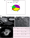
Comparative analysis of third-generation dual-energy CT and IVUS for in-stent restenosis detection
- PMID: 40835906
- PMCID: PMC12366176
- DOI: 10.1186/s12872-025-04836-z
Comparative analysis of third-generation dual-energy CT and IVUS for in-stent restenosis detection
Abstract
Purpose: Prior studies have assessed in-stent diameter restenosis (ISDR) in coronary arteries using 64-slice multidetector computed tomography coronary angiography (MDCT-CA) compared to invasive coronary angiography (ICA), which is the gold standard. This study aimed to compare the diagnostic accuracy of monoenergetic reconstruction using third-generation dual-source dual-energy CT (DSDECT) to that of ICA reconstruction via adjunctive intravascular ultrasonography (IVUS) for evaluating the ISDR.
Methods: A total of 95 patients with previously stented coronary arteries (involving 110 stents) underwent DSDECT followed by ICA and IVUS within a 24-h timeframe. The specificities, sensitivities, negative predictive values (NPVs), and positive predictive values (PPVs) of the DSDECT and ICA were compared for confirming or excluding the ISDR using in-stent area restenosis (ISAR) and a minimal luminal area (MLA) ≤ 4.0 mm2 on IVUS as the reference standard.
Results: Compared with IVUS, the latest DSDECT demonstrated good sensitivity (100%), specificity (92.4%), and accuracy (96.1%) in detecting the ISDR. Our study highlights a limitation in assessability for stents with diameters < 3 mm, emphasizing the importance of careful patient selection. When employing an IVUS MLA of 4.0 mm2 as a reference for identifying the ISDR, no significant difference was observed between DSDECT and ICA in the identification of the ISDR. However, it is important to note that the use of absolute cut-offs, such as < 6.0 mm2 in the left main or < 4.0 mm2, may not universally apply across varying ethnicities and between sexes. The interpretation of the minimal luminal area (MLA) should be considered in the context of individual patient characteristics, and caution is advised to avoid potential misleading conclusions based solely on absolute thresholds.
Conclusion: In summary, when assessing stent patency, the latest DSDECT exhibits similar performance to coronary angiography and IVUS. Moreover, it offers noninvasiveness, cost-effectiveness, and ease of operation, which are advantageous characteristics. However, it is essential to consider limitations in patient eligibility, including factors such as prior cardiac devices, arrhythmias, and any degree of chronic renal insufficiency, which may impact CT imaging analysis. The 100% negative predictive value (NPV) of third-generation DSDECT reliably excludes in-stent restenosis (ISDR), potentially obviating invasive angiography in stable patients with patent stents.
Trial registration: ZU-IRB#3915/13-8-2017 Registered 13 August 2017, email: IRB_123@medicine.zu.edu.eg.
Keywords: Computed tomography angiography; Coronary atherosclerosis; Coronary intervention planning; In-stent restenosis; Intravascular ultrasound; Noninvasive imaging.
© 2025. The Author(s).
Conflict of interest statement
Declarations. Ethics approval and consent to participate: The protocol was approved by the Institutional Review Board (IRB), Faculty of Medicine, Zagazig University (ZU-IRB#3915/13–8-2017), which confirmed that all procedures were performed in accordance with the ethical standards of the institutional and/or national research committee and with the 1964 Helsinki Declaration and its later amendments or comparable ethical standards. Consent for publication: We confirm that informed consent was obtained from all study participants or their legal guardians for the publication of identifying information or images in this online open-access publication. All efforts were made to ensure the privacy and confidentiality of the participants involved in this study. Competing interests: The authors declare no competing interests.
Figures


Similar articles
-
Evaluation of coronary artery stent patency by using 64-slice multi-detector computed tomography and conventional coronary angiography: a comparison with intravascular ultrasonography.Int J Cardiol. 2013 Jun 5;166(1):90-5. doi: 10.1016/j.ijcard.2011.10.003. Epub 2011 Nov 5. Int J Cardiol. 2013. PMID: 22056475
-
Systematic review of the clinical effectiveness and cost-effectiveness of 64-slice or higher computed tomography angiography as an alternative to invasive coronary angiography in the investigation of coronary artery disease.Health Technol Assess. 2008 May;12(17):iii-iv, ix-143. doi: 10.3310/hta12170. Health Technol Assess. 2008. PMID: 18462576
-
Prescription of Controlled Substances: Benefits and Risks.2025 Jul 6. In: StatPearls [Internet]. Treasure Island (FL): StatPearls Publishing; 2025 Jan–. 2025 Jul 6. In: StatPearls [Internet]. Treasure Island (FL): StatPearls Publishing; 2025 Jan–. PMID: 30726003 Free Books & Documents.
-
Diagnostic accuracy of dual-source computed tomography angiography for the detection of coronary in-stent restenosis: A systematic review and meta-analysis.Echocardiography. 2018 Apr;35(4):541-550. doi: 10.1111/echo.13863. Epub 2018 Mar 23. Echocardiography. 2018. PMID: 29569751
-
Intravascular ultrasound-guided interventions in coronary artery disease: a systematic literature review, with decision-analytic modelling, of outcomes and cost-effectiveness.Health Technol Assess. 2000;4(35):1-117. Health Technol Assess. 2000. PMID: 11109031
References
-
- Knuuti J, Wijns W, Saraste A, et al. 2019 ESC Guidelines for the diagnosis and management of chronic coronary syndromes. Eur Heart J. 2020;41(3):407–77. 10.1093/eurheartj/ehz425. - PubMed
-
- Achenbach S. Computed tomography coronary angiography. J Am Coll Cardiol. 2006;48:1919–28. 10.1016/j.jacc.2006.08.025. - PubMed
-
- Schuijf JD, Bax JJ, Jukema JW. Feasibility of assessment of coronary stent patency using 16-slice computed tomography. Am J Cardiol. 2004;94:427–30. 10.1016/j.amjcard.2004.05.045. - PubMed
-
- Mark DB, Berman DS, Budoff MJ, et al. ACCF/ACR/AHA/NASCI/SAIP/SCAI/SCCT 2010 expert consensus document on coronary computed tomographic angiography: a report of the American College of Cardiology Foundation Task Force on Expert Consensus Documents. J Am Coll Cardiol. 2010;55:2663–99. 10.1016/j.jacc.2010.02.013. - PubMed
-
- Rixe J, Achenbach S, Ropers D, et al. Assessment of coronary artery stent restenosis by 64-slice multidetector computed tomography. Eur Heart J. 2006;27:2567–72. 10.1093/eurheartj/ehl296. - PubMed
Publication types
MeSH terms
LinkOut - more resources
Full Text Sources
Medical
Miscellaneous

