Multicenter Study
. 2025 Nov;31(11):3832-3844.
doi: 10.1038/s41591-025-03916-z. Epub 2025 Aug 20.

AI-based diagnosis of acute aortic syndrome from noncontrast CT

Affiliations
Multicenter Study

AI-based diagnosis of acute aortic syndrome from noncontrast CT

Yujian Hu et al. Nat Med. 2025 Nov.

Abstract

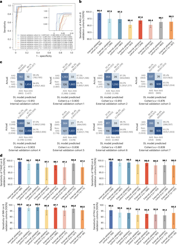
The accurate and timely diagnosis of acute aortic syndrome (AAS) in patients presenting with acute chest pain remains a clinical challenge. Aortic computed tomography (CT) angiography is the imaging protocol of choice in patients with suspected AAS. However, due to economic and workflow constraints in China, the majority of suspected patients initially undergo noncontrast CT as the initial imaging testing, and CT angiography is reserved for those at higher risk. Although noncontrast CT can reveal specific signs indicative of AAS, its diagnostic efficacy when used alone has not been well characterized. Here we present an artificial intelligence-based warning system, iAorta, using noncontrast CT for AAS identification in China, which demonstrates remarkably high accuracy and provides clinicians with interpretable warnings. iAorta was evaluated through a comprehensive step-wise study. In the multicenter retrospective study (n = 20,750), iAorta achieved a mean area under the receiver operating curve of 0.958 (95% confidence interval 0.950-0.967). In the large-scale real-world study (n = 137,525), iAorta demonstrated consistently high performance across various noncontrast CT protocols, achieving a sensitivity of 0.913-0.942 and a specificity of 0.991-0.993. In the prospective comparative study (n = 13,846), iAorta demonstrated the capability to significantly shorten the time to correct diagnostic pathway for patients with initial false suspicion from an average of 219.7 (115-325) min to 61.6 (43-89) min. Furthermore, for the prospective pilot deployment that we conducted, iAorta correctly identified 21 out of 22 patients with AAS among 15,584 consecutive patients presenting with acute chest pain and under noncontrast CT protocol in the emergency department. For these 21 AAS-positive patients, the average time to diagnosis was 102.1 (75-133) min. Finally, iAorta may help prevent delayed or missed diagnoses of AAS in settings where noncontrast CT remains the only feasible initial imaging modality-such as in resource-limited regions or in patients who cannot receive, or did not receive, intravenous contrast.


Conflict of interest statement

Competing interests: Alibaba Group has filed for patent protection (application number CN 202311181343.8) on behalf of Y.-J.Z., M.X. and L. Lu for the work related to the methods of detection of acute aortic syndrome on noncontrast CT. Y.-J.Z., W.G., J.Z., T.C.W.M., Z.L., L.L. and M.X. are employees of Alibaba Group and own Alibaba stock as part of the standard compensation package. All other authors have no competing interests.

Figures

Fig. 1
Fig. 1. Overall study design and model pipeline.
a, Clinical starting point for the study. The diagnosis of AAS poses a notable challenge within the ED owing to its nonspecific clinical symptoms. In China, more than half of the patients with acute chest pain are initially suspected of less critical illnesses and thus received noncontrast CT scans as the initial imaging test. Our goal is to develop an AI model that can rapidly and accurately identify patients with suspected AAS from this population of individuals undergoing noncontrast CT scans, while providing interpretable results to assist radiologists and physicians in making informed clinical decisions. b, A schematic overview of the model. It was trained with patient-level diagnostic labels and segmentation masks annotated on arterial phase series. The model takes noncontrast phase series as input and outputs the probability of AAS, segmentation masks of the aortic wall and true lumen, and an activation map highlighting potential lesion areas. c, Retrospective and prospective evaluation of model and iAorta. Retrospective evaluation for model consists of multicenter model validation (stage I, n = 20,750), reader study (stage II, n = 2,287) and large-scale real-world study (stage III, n = 137,525). Prospective evaluation (stage IV) for iAorta consists of comparative study (n = 13,846) and pilot deployment study (n = 15,584). iAorta incorporates a phase selection module, our model and a pop-up warning module.
Fig. 2
Fig. 2. Stage I multicenter model validation.
a, ROC curves of AAS detection on the internal and external validation cohorts. b, Sensitivity of AAS detection in the internal validation cohort (n = 795) and external validation cohorts (cohorts 1–7, n = 6,495). The error bars denote the two-sided 95% CI computed from 1,000 bootstrapping iterations. c, Confusion matrices of AAS detection on the internal and external validation cohorts showing TPs, TNs, FPs, and FNs, with sensitivity, specificity, PPV and NPV calculated accordingly. d, Sensitivity of four subtypes (TAAD, TBAD, IMH and PAU) of AAS detection in the internal cohorts (n = 795) and external validation cohorts (cohorts 1–7, n = 6,495). The error bars denote the two-sided 95% CI computed from 1,000 bootstrapping iterations. FPs, false positives; FNs, false negatives; TPs, true positives; TNs, true negatives.
Fig. 3
Fig. 3. Stage II reader study.
a, Comparison between the DL model and 11 radiologists with different levels of expertise on noncontrast CT for AAS detection, with or without the assistance of model. b, Sensitivity of 11 radiologists with different levels of expertise on noncontrast CT for four different subtypes of AAS detection, with or without the assistance of model (n = 795 data samples). On each box in b, the central line indicates the median, and the bottom and top edges of the box indicate the 25th and 75th percentiles, respectively. The whiskers extend to 1.5 times the interquartile range. c, Examples of the ‘visible’ positive case of TBAD, ‘invisible’ positive case of IMH and negative case with an ascending aorta artifact, which was not detected by readers on noncontrast CT but well classified with the assistance of the model.
Fig. 4
Fig. 4. Stage III large-scale real-world retrospective study.
a, The patients’ distribution in the study. b, The distributions of CT protocols in the study. c, The sensitivity and specificity of original model on RW1 (n = 23,094) and that of original and upgraded model on RW2 (cohorts 1–3, n = 122,107). The superscript asterisk represents results of the upgraded model. The error bars denote the two-sided 95% CI computed from 1,000 bootstrapping iterations. d, ROC curves of AAS detection on the RW1 and RW2 cohorts. Blue and orange curves represent the performance of the original model and the upgraded model, respectively. e, Model performance metrics (AUC, accuracy, sensitivity and specificity) for AAS detection across three subtypes (noncontrast chest CT (cohorts 1–3, n = 42,423), noncontrast abdominal CT (cohorts 1–3, n = 74,261) and others (cohorts 1–3, n = 5,423)) in the RW2 cohorts. The error bars denote the two-sided 95% CI computed from 1,000 bootstrapping iterations. SEN, sensitivity; SPE, specificity.
Fig. 5
Fig. 5. Stage IV prospective multicenter study (comparative study).
a, The study involves two groups of radiologists: group A, the radiologist team, consisting of the IR radiologist and the RR radiologist, who independently evaluate images in a sequential manner, mirroring the current conventional clinical workflow; and group B, the radiologist team evaluating images assisted by the iAorta system. If iAorta detects any abnormalities, both the IR and RR radiologists receive sequential pop-up alerts, prompting them to prioritize the review of the AI-flagged AAS-positive image. RIS, radiology information system. b, The sensitivity and specificity of group A (IR radiologist and RR radiologist), group B (IR radiologist + iAorta and RR radiologist + iAorta) and iAorta system (n = 14,436 data samples). The error bars denote the two-sided 95% CI computed from 1,000 bootstrapping iterations. The P value was calculated by two-sided McNamar test (P < 0.001 (3.47 × 10−21)). c, Time from presentation to correct diagnostic pathway (group A versus group B) in the realistic clinical settings. Nine patients with AAS were identified by iAorta, yet two patients with AAS (PAU) were missed. d, An illustrative example highlights the potential benefits of using iAorta for patients with AAS who initially were suspected of having other acute conditions and underwent noncontrast CT in real-world emergency settings.iAorta has demonstrated the potential to significantly reduce the time needed to reach a correct diagnosis for this patient, from 273 min to 56 min.
Fig. 6
Fig. 6. Stage IV prospective multicenter study (pilot deployment study).
a, iAorta is seamlessly incorporated into the existing pilot clinical routine. Once iAorta identifies patients at risk of AAS, it generates pop-up alerts for the radiology team, prompting them to prioritize the review of the flagged images. b, Confusion matrices of AAS detection in this study showing the TPs, TNs, FPs and FNs of AAS detection and the sensitivity (95.5%), specificity (99.4%), PPV (18.6%) and NPV (99.9%) calculated accordingly. c, Potential benefits of the iAorta system in the realistic clinical settings of China. d, An illustrative example highlighting the benefits of using iAorta for patients with AAS who initially presented with acute abdominal pain and were suspected of having other acute conditions in real-world emergency settings. While the patient initially entered an incorrect diagnostic pathway, the early warning provided by iAorta on the noncontrast CT enabled the definitive diagnosis of AAS within 94 min of hospital admission, ensuring both timely and appropriate management. FPs, false positives; FNs, false negatives; TPs, true positives; TNs, true negatives; BP, blood pressure; HR, heart rate; RR, respiratory rate; CRP, C-reactive protein.
Extended Data Fig. 1
Extended Data Fig. 1. Patient and data sources of model development, multi-center model validation (stage I), reader study (stage II), large-scale real-world retrospective study (stage III), and prospective multi-center study (stage IV).
Data source includes different field of views (FOVs) and z-axis coverage in aortic CTA, coronary CTA, non-contrast chest CT, pulmonary CTA, rib CT, non-contrast abdominal CT, and lumbar spine CT.
Extended Data Fig. 2
Extended Data Fig. 2. Network architecture.
(Top) Overview. Our deep learning framework consists of two stages: aorta localization using a lightweight U-Net, and abnormality detection using a multi-task CNN. (Bottom) Architectures of the lightweight U-Net and multi-task CNN. The features extracted from encoder are used for abnormal and normal classification. Decoder A and T are designed for the segmentation of aorta and true lumen, respectively.
Extended Data Fig. 3
Extended Data Fig. 3. Interpretable results of iAorta in four subtypes of AAS and two artifact cases.
The two colors in the slice sequence (arranged by spatial location) represent two classes of slice-level prediction including AAS (yellow) and non-AAS (green). The slice activation map indicates the voxel-level localization of AAS. AAS, acute aortic syndrome; TAAD, Stanford Type A dissection; TBAD, Stanford Type B dissection; IMH, intramural hematoma; PAU, penetrating atherosclerotic ulcer.
Extended Data Fig. 4
Extended Data Fig. 4. Flowchart illustrating the potential benefits for patients with Acute Aortic Syndrome (AAS) who were initially suspected to have other acute diseases in the retrospective real-world study (stage III), compared to the current clinical workflow.
a. A patient with lower back pain was initially suspected to have lumbar disc protrusion and underwent clinical investigations, including lumbar spine CT and non-contrast chest CT. b. A patient with abdominal pain was initially suspected to have acute gastroenteritis, underwent clinical investigations including a non-contrast abdominal CT, and was discharged from the ED. Four days later, he returned to the ED with worsening symptoms and underwent a non-contrast chest CT. Our model could have detected AAS during the first visit. c. A patient with abdominal pain was suspected to have a chemotherapy drug reaction and underwent clinical investigations, including non-contrast abdominal CT and pulmonary CTA. Before a definitive diagnosis, the patient experienced cardiopulmonary arrest. iAorta system could potentially save the patient’s life.
Extended Data Fig. 5
Extended Data Fig. 5. Flowchart describing the process of the seamless integration of iAorta into the existing clinical workflow.
PACS, picture archiving and communication system; RIS, radiology information system.

References

    1. Isselbacher, E. M. et al. 2022 ACC/AHA Guideline for the Diagnosis and Management of Aortic Disease. J. Am. Coll. Cardiol.80, e223–e393 (2022). - PMC - PubMed
    1. Mazzolai, L. et al. 2024 ESC guidelines for the management of peripheral arterial and aortic diseases. Eur. Heart J.45, 3538–3700 (2024). - PubMed
    1. Hagan, P. G. et al. The International Registry of Acute Aortic Dissection (IRAD): new insights into an old disease. JAMA283, 897–903 (2000). - PubMed
    1. Strayer, R. J. et al. Screening, evaluation, and early management of acute aortic dissection in the ED. Curr. Cardiol. Rev.8, 152–157 (2012). - PMC - PubMed
    1. Harris, K. M. et al. Correlates of delayed recognition and treatment of acute type A aortic dissection: the IRAD experience. Circulation124, 1911–1918 (2011). - PubMed

Publication types