. 2025 Aug 20;15(1):30552.
doi: 10.1038/s41598-025-13309-4.

Adipocyte-derived factors induce adherent to suspension transition in breast and pancreatic cancer cells through lipid metabolic alteration

Affiliations

Adipocyte-derived factors induce adherent to suspension transition in breast and pancreatic cancer cells through lipid metabolic alteration

Doru Kwon et al. Sci Rep. .

Abstract

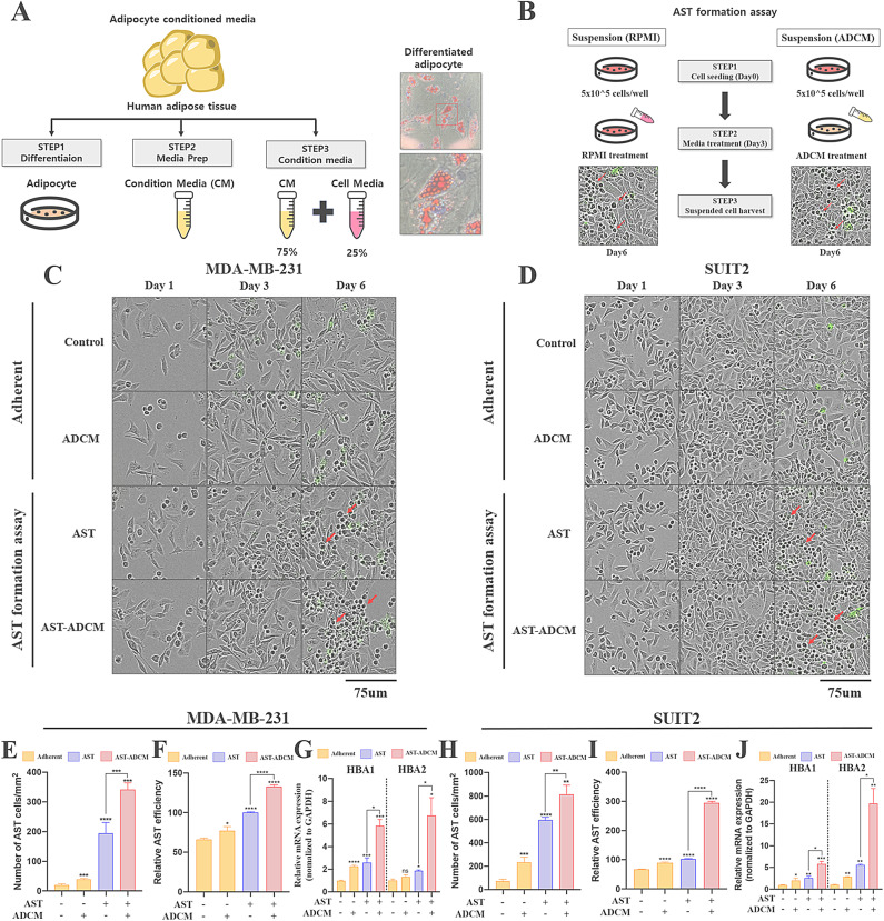
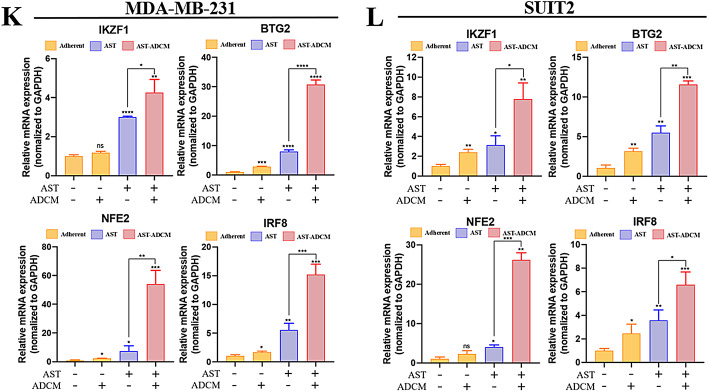
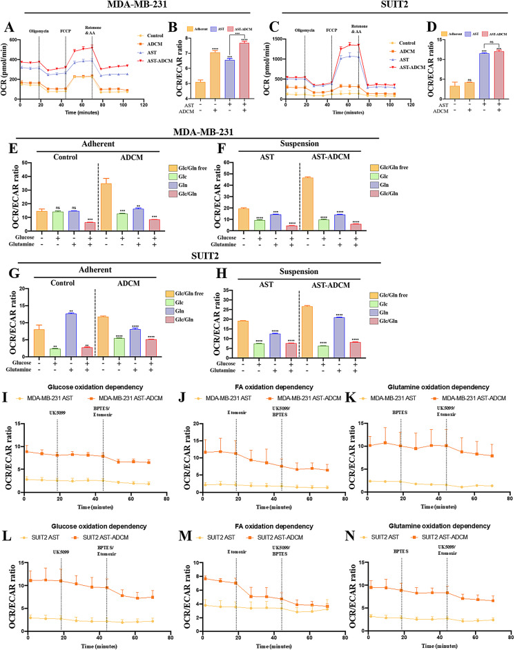
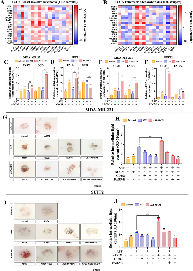
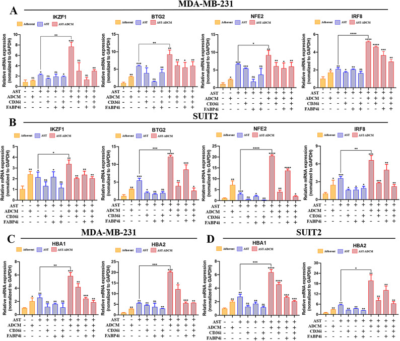
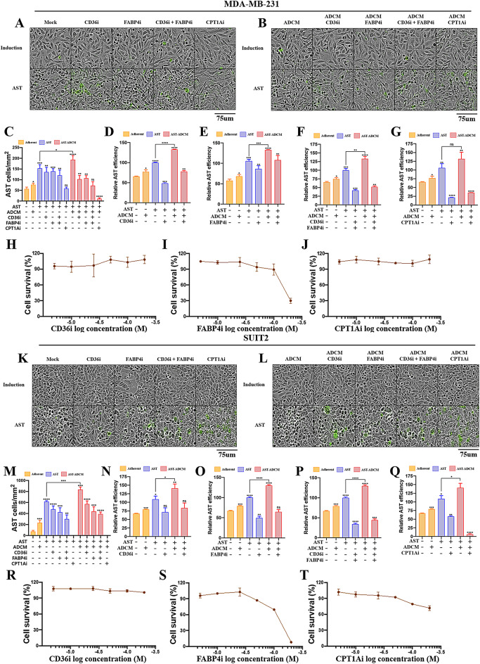
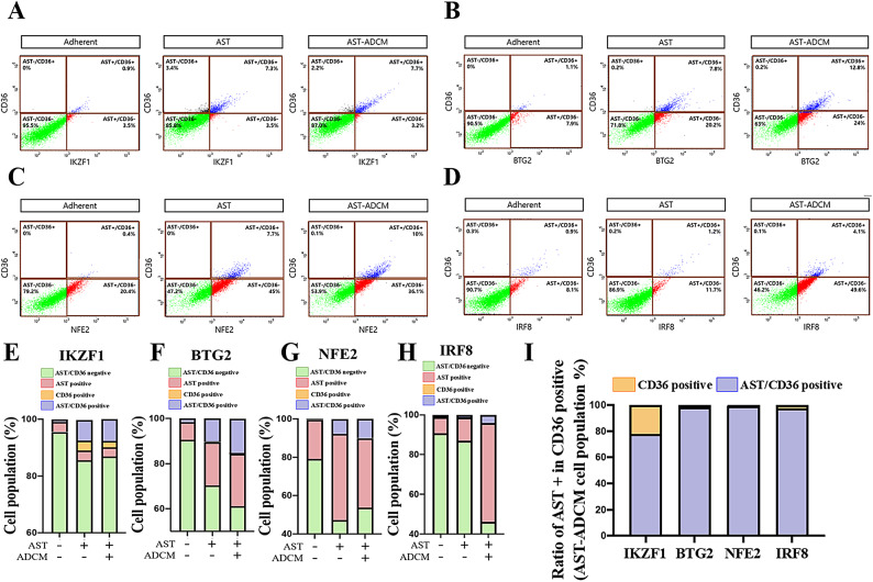
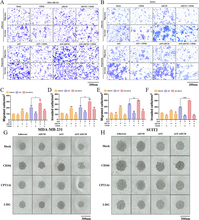
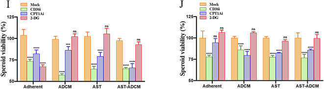
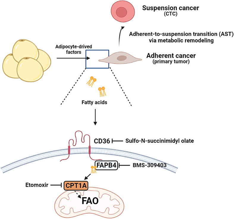
Adipocytes play a dynamic role in the tumor microenvironment (TME) by acting as facilitators, providing cytokines and metabolites that regulate cancer progression and metastasis. Despite metastasis being a major contributor to cancer-associated mortality, our understanding of how adipocytes influence this process remains limited. This study aims to elucidate the regulatory mechanism of Adherent to Suspension Transition (AST) reprogramming within the adipocyte, driven by anchorage dependency. AST facilitates the conversion of adherent tumor cells into suspension cells, thereby contributing to the generation of circulating tumor cells (CTCs). We have evaluated generating AST cells from primary tumors using a dissemination assay that mimics CTCs in vitro. Additionally, we examined AST cell formation when incubated with human adipocyte-conditioned media (ADCM) using the InCucyte live-cell imaging system. Through this approach, we effectively assessed the impact of the tumor-adipocyte interactions on CTC formation from the perspective of AST. As a metastasis-initiating marker, CD36 is pivotal in fatty acid (FA) acquisition and regulates lipid metabolic remodeling during the AST. The generation of AST cells through AST reprogramming is controlled by fatty acid oxidation (FAO), and pharmacological blockade of CD36 and FAO significantly reduced AST cell generation. This demonstrates that CD36 plays a key role in the early stages of AST-induced dissemination. Additionally, promoting cancer cell aggressiveness through ADCM enhances metastatic potency and upregulates the expression of AST reprogramming factors. Inhibition of lipid metabolism not only suppresses AST cell formation but also decreases survival in suspension. This indicates that exogenous lipid uptake and FAO via CD36 play crucial roles in the metastasis process, facilitating the dissemination of primary tumors into the bloodstream. Adipocytes contribute to cancer progression by supplying various metabolites to cancer cells. While primary tumors predominantly rely on glucose as a major energy source, cellular remodeling during dissemination shifts metabolic dependency toward lipids. In the TME, where adipocytes are abundant, tumor cells acquire FA through CD36-mediated uptake for metabolic adaptation. This shift to lipid metabolism is essential for AST, and thus, targeting lipid metabolism via inhibition of CD36 and FAO could serve as a potential therapeutic strategy for AST.

Keywords: AST; Adipocytes; CD36; Circulating tumor cell; Lipid metabolism; Tumor microenvironment.


Conflict of interest statement

Declarations. Ethics approval and consent to participate: Human adipose tissue usage received ethical approval from the Institutional Review Board at Gangnam Severance Hospital, Yonsei University, Seoul, Republic of Korea, adhering to the ethical principles outlined in the 1975 Declaration of Helsinki. Acquisition of human adipose tissue samples was conducted with the informed consent of participating patients, following guidelines and regulations approved by the Institutional Review Board of Gangnam Severance Hospital, Yonsei University Health System. Consent for publication: All authors have consented to their inclusion in the list and have approved the submission of this content. The material has not been presented for publication elsewhere during the review process for consideration. Competing interests: The authors declare no competing interests.

Figures

Fig. 1
Fig. 1
Adipocyte-conditioned media promote adherent to suspension transition in MDA-MB-231 and SUIT2. (A) A schematic representation of ADCM extraction from human adipose tissue. Pre-adipocytes were isolated from human adipose tissue and differentiated into mature adipocytes. ADCM was used at a working concentration of 75% ADCM mixed with 25% complete RPMI 1640 medium for the dissemination assay. Illustration was created using BioRender (BioRender.com). (B) A diagram of dissemination assay and incubation with ADCM. After three days of culture, AST induction was performed using complete media or ADCM, respectively. Cells were harvested after 72 h of induction. (C, D) Live cell images of MDA-MB-231 and SUIT2 at different time points (days 1, 3, and 6). Red arrows indicated the AST cells. (E, H) Quantification of AST cells after cell dissemination assay in MDA-MB-231 and SUIT2 cultured with or without ADCM. (F, I) Quantification of AST efficiency of MDA-MB-231 and SUIT2 cultured with or without ADCM. Efficiency was measured by the CellTiter Glo® 2.0 cell viability assay after 2 days of culture in an ultra-low binding plate. (G, J) qRT-PCR was used to examine HBA1 and HBA2 expression in cell culture with or without ADCM and dissemination assay. (K, L) Quantifying mRNA expression levels of 4 key AST factors in MDA-MB-231 and SUIT2 culture with or without ADCM and dissemination assay. Relative mRNA expression was normalized to GAPDH. Data are presented as mean ± SD. Statistical significance was determined using Student’s t-test. ****p < 0.0001; ***p < 0.001; **p < 0.01; *p < 0.05. All statistical comparisons were made against the control (adherent cells without ADCM). All images were acquired with magnification 10X, scale bar = 75 μm.
Fig. 1
Fig. 1
Adipocyte-conditioned media promote adherent to suspension transition in MDA-MB-231 and SUIT2. (A) A schematic representation of ADCM extraction from human adipose tissue. Pre-adipocytes were isolated from human adipose tissue and differentiated into mature adipocytes. ADCM was used at a working concentration of 75% ADCM mixed with 25% complete RPMI 1640 medium for the dissemination assay. Illustration was created using BioRender (BioRender.com). (B) A diagram of dissemination assay and incubation with ADCM. After three days of culture, AST induction was performed using complete media or ADCM, respectively. Cells were harvested after 72 h of induction. (C, D) Live cell images of MDA-MB-231 and SUIT2 at different time points (days 1, 3, and 6). Red arrows indicated the AST cells. (E, H) Quantification of AST cells after cell dissemination assay in MDA-MB-231 and SUIT2 cultured with or without ADCM. (F, I) Quantification of AST efficiency of MDA-MB-231 and SUIT2 cultured with or without ADCM. Efficiency was measured by the CellTiter Glo® 2.0 cell viability assay after 2 days of culture in an ultra-low binding plate. (G, J) qRT-PCR was used to examine HBA1 and HBA2 expression in cell culture with or without ADCM and dissemination assay. (K, L) Quantifying mRNA expression levels of 4 key AST factors in MDA-MB-231 and SUIT2 culture with or without ADCM and dissemination assay. Relative mRNA expression was normalized to GAPDH. Data are presented as mean ± SD. Statistical significance was determined using Student’s t-test. ****p < 0.0001; ***p < 0.001; **p < 0.01; *p < 0.05. All statistical comparisons were made against the control (adherent cells without ADCM). All images were acquired with magnification 10X, scale bar = 75 μm.
Fig. 2
Fig. 2
AST cells showed increased oxygen consumption via lipid metabolic alterations in MDA-MB-231 and SUIT2. (A, C) The XFe24 seahorse oxygen consumption assay was carried out in MDA-MB-231 (A) and SUIT2 (C) cells with or without ADCM and dissemination assay. Each group was injected with the stressors oligomycin, FCCP, and rotenone/antimycin A (Rotenone/AA) into the media. (B, D) The ratio of OCR to ECAR was measured in MDA-MB-231 and SUIT2 cells. (E ~ H) The OCR/ECAR ratio alteration was assessed in both MDA-MB-231 and SUIT2 cells in both adherent and AST states. Values were measured upon individual or combined glucose and glutamine deprivation. Experiments were conducted with four groups: Glucose+/Glutamine+ (Glc+/Gln+), Glucose+ (Glc+), Glutamine+ (Gln+), and nutrient-free condition. (I ~ N) The XF Mito Fuel Flex test was performed in MDA-MB-231 and SUIT2 cells cultured with or without ADCM and subjected to a dissemination assay. Each group was sequentially treated with specific metabolic inhibitors: UK5099 (glucose metabolism inhibitor), Etomoxir (fatty acid oxidation inhibitor), and BPTES (glutamine metabolism inhibitor). To assess the dependency on each metabolic pathway, OCR and ECAR were first measured following the addition of each inhibitor alone. Subsequent changes in OCR and ECAR upon treatment with the remaining two inhibitors were then analyzed to evaluate the cells’ metabolic sensitivity. Data are presented as mean ± SD. Statistical significance was determined using Student’s t-test. ****p < 0.0001; ***p < 0.001; **p < 0.01; *p < 0.05. All statistical comparisons were made against the control group.
Fig. 3
Fig. 3
An up-regulated lipid uptake pathway increases lipid accumulation in MDA-MB-231 and SUIT2. (A, B) TCGA micro-array mRNA expression data for breast invasive carcinoma (1108 samples in A) and pancreatic adenocarcinoma (186 samples in B) were used. Comparison was conducted between AST factors and a gene set related to lipid transport. Spearman’s correlation was measured by comparing the data. (C, D) The mRNA levels of FASN and SCD involved in the endogenous lipid synthesis pathway were measured in MDA-MB-231 (C) and SUIT2 (D) cultures with or without ADCM and dissemination assay. (E, F) The mRNA levels of CD36 and FABP4 involved in the exogenous lipid uptake pathway were measured in MDA-MB-231 (E) and SUIT2 (F) cultures with or without ADCM and dissemination assay. (G ~ J) Oil-Red O staining was used to determine MDA-MB-231 and SUIT2 lipid accumulation. Sulfo-N-succinimidyl olate, a CD36 inhibitor, and BMS-309403, a FABP4 inhibitor, were used to inhibit exogenous lipid uptake in MDA-MB-231 (G, H) and SUIT2 (I, J) culture with or without ADCM and dissemination assay. Relative mRNA expression was normalized to GAPDH. Data are presented as mean ± SD. Statistical significance was determined using Student’s t-test. ****p < 0.0001; ***p < 0.001; **p < 0.01; *p < 0.05. All statistical comparisons were made against the control (adherent cells without ADCM). All images were acquired with magnification 40X, scale bar = 10 μm.
Fig. 4
Fig. 4
Sulfo-N-Succinimidyl Olate and BMS-309403 decrease adherent to suspension transition factors in MDA-MB-231 and SUIT2. (A ~ D) The CD36 inhibitor Sulfo-N-Succinimidyl Olate and the FABP4 inhibitor BMS-309403 were used to compare four key AST factor expression and hematopoietic genes in MDA-MB-231 (A, C) and SUIT2 (B, D) cells based on exogenous lipid uptake inhibition. Relative mRNA expression was normalized to GAPDH. Data are presented as mean ± SD. Statistical significance was determined using Student’s t-test. ****p < 0.0001; ***p < 0.001; **p < 0.01; *p < 0.05. All statistical comparisons were made against the control (adherent cells without ADCM).
Fig. 5
Fig. 5
Inhibition of fatty acid oxidation decreases adherent to suspension transition. (A ~ C, K ~ M) MDA-MB-231 (A ~ C) and SUIT2 (K ~ M) AST cell formations were observed when treated with CD36, FABP4, and CPT1A inhibitors in culture with or without ADCM and dissemination assay. (D ~ G, N ~ Q) AST efficiency was assessed in MDA-MB-231 (D ~ G) and SUIT2 (N ~ Q) culture with or without ADCM and dissemination assay when treated with CD36 inhibitor (D, N), FABP4 inhibitor (E, O), CD36 and FABP4 inhibitor (F, P), and CPT1A inhibitor (G, Q). (H ~ J, R ~ T) Cell viability measurements in MDA-MB-231 (H ~ J) and SUIT2 (R ~ T) cells after treatment with CD36 inhibitor (H, R), FABP4 inhibitor (I, S), and CPT1A inhibitor (J, T). Data are presented as mean ± SD. Statistical significance was determined using Student’s t-test. ****p < 0.0001; ***p < 0.001; **p < 0.01; *p < 0.05. All statistical comparisons were made against the control (adherent cells without ADCM). All images were acquired with magnification 10X, scale bar = 75 μm.
Fig. 6
Fig. 6
CD36 has a positive correlation with four key adherent to suspension transition factors. (A ~H) Flow cytometry was performed to compare CD36 to AST factor expression in SUIT2 cell culture with or without ADCM and dissemination assay. They were separated and measured into AST - / CD36 -, AST + / CD36 -, AST - / CD36 +, and AST + / CD36 + groups based on the AST factor and CD36 expression. (I) Quantitative measurement of the AST + /CD36 + cells in total CD36 + cells according to the AST cells with ADCM.
Fig. 7
Fig. 7
Inhibition of fatty acid uptake leads to a reduction in the metastatic ability of AST. (A, B) Migration and invasion were measured in adherent and AST cells of MDA-MB-231 (A) and SUIT2 (B) when the CD36 inhibitor was treated. (C ~ F) MDA-MB-231 (C, D) and SUIT2 (E, F) migrated and invaded cells after being treated with CD36 inhibitor were quantified. (G, H) Spheroid formation assay was performed in MDA-MB-231 (G) and SUIT2 (H) cells. To assess the impact of metabolism inhibitors on spheroid cluster formation, CD36i, CPT1Ai, and 2-DG were individually administered. (I, J) Measurement of spheroid viability in MDA-MB-231 (I) and SUIT2 (J) cells upon treatment with metabolism inhibitors. Data are presented as mean ± SD. Statistical significance was determined using Student’s t-test. ****p < 0.0001; ***p < 0.001; **p < 0.01; *p < 0.05. All statistical comparisons were made against the control. A, B were acquired with magnification 5 X, scale bar = 200 μm. G, H were acquired with magnification 10 X, scale bar = 300 μm.
Fig. 7
Fig. 7
Inhibition of fatty acid uptake leads to a reduction in the metastatic ability of AST. (A, B) Migration and invasion were measured in adherent and AST cells of MDA-MB-231 (A) and SUIT2 (B) when the CD36 inhibitor was treated. (C ~ F) MDA-MB-231 (C, D) and SUIT2 (E, F) migrated and invaded cells after being treated with CD36 inhibitor were quantified. (G, H) Spheroid formation assay was performed in MDA-MB-231 (G) and SUIT2 (H) cells. To assess the impact of metabolism inhibitors on spheroid cluster formation, CD36i, CPT1Ai, and 2-DG were individually administered. (I, J) Measurement of spheroid viability in MDA-MB-231 (I) and SUIT2 (J) cells upon treatment with metabolism inhibitors. Data are presented as mean ± SD. Statistical significance was determined using Student’s t-test. ****p < 0.0001; ***p < 0.001; **p < 0.01; *p < 0.05. All statistical comparisons were made against the control. A, B were acquired with magnification 5 X, scale bar = 200 μm. G, H were acquired with magnification 10 X, scale bar = 300 μm.
Fig. 8
Fig. 8
Pharmacological targeting of lipid metabolism inhibits adherent to suspension transition. FA supplied by adipocytes is absorbed by cancer cells via the CD36 transporter. The FA delivered into mitochondria through CPT1A contributes to fulfilling the metabolic requirements of cancer cells through fatty acid oxidation. The increase in lipid metabolism through lipid metabolic alterations triggers the adherent to suspension transition. This transition can be effectively inhibited by antagonists targeting CD36, FABP4, and CPT1A, which are involved in this process. Illustration was created using BioRender (BioRender.com).

Similar articles

References

    1. Dillekås, H., Rogers, M. S. & Straume, O. Are 90% of deaths from cancer caused by metastases? Cancer Med.8, 5574–5576. 10.1002/cam4.2474 (2019). - PMC - PubMed
    1. Swartz, M. A. et al. Tumor microenvironment complexity: emerging roles in cancer therapy. Cancer Res.72, 2473–2480. 10.1158/0008-5472.Can-12-0122 (2012). - PMC - PubMed
    1. Joyce, J. A. & Pollard, J. W. Microenvironmental regulation of metastasis. Nat. Rev. Cancer. 9, 239–252. 10.1038/nrc2618 (2009). - PMC - PubMed
    1. Mukherjee, A., Bilecz, A. J. & Lengyel, E. The adipocyte microenvironment and cancer. Cancer Metastasis Rev.41, 575–587. 10.1007/s10555-022-10059-x (2022). - PubMed
    1. Wang, Z. et al. Metastasis-associated fibroblasts: an emerging target for metastatic cancer. Biomark. Res.910.1186/s40364-021-00305-9 (2021). - PMC - PubMed