. 2025 Aug 20;25(1):605.
doi: 10.1186/s12876-025-03978-w.

Hypoxia-induced exosomal circNRIP1 activates cancer-associated fibroblasts to promote esophageal squamous cell carcinoma migration and invasion

Affiliations

Hypoxia-induced exosomal circNRIP1 activates cancer-associated fibroblasts to promote esophageal squamous cell carcinoma migration and invasion

Guan'en Qiao et al. BMC Gastroenterol. .

Abstract

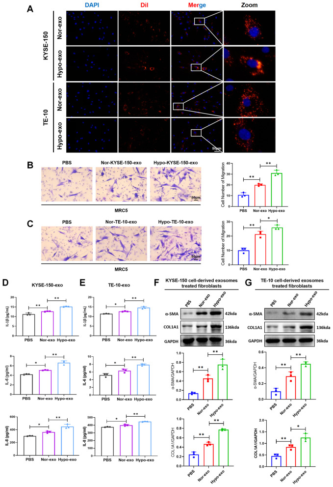
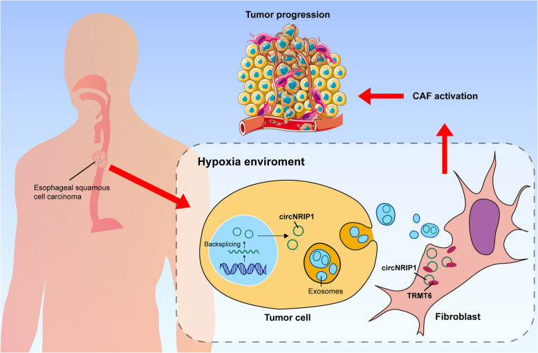
Esophageal squamous cell carcinoma (ESCC) is characterized by a complex tumor microenvironment (TME). Cancer-associated fibroblasts (CAFs) play a crucial role in the TME that facilitate tumor progression via interactions with cancer cells. However, the mechanisms underlying the activation of CAFs in TME remain largely unknown. Here, we characterized the exosomes derived from normoxic and hypoxic ESCC cells using electron microscopy and western blot. The impact of exosomes on CAF activation and the motility of ESCC cells was examined in vitro. The molecular complex involving circNRIP1 was explored using RNA pull-down. We demonstrated that exosomes derived from ESCC cells, including KYSE-150 and TE-10 cells, exhibited a significantly increase in secretion under hypoxic conditions. These hypoxic exosomes were internalized by fibroblasts and further promoted the transformation of normal fibroblasts into CAFs, as evidenced by enhanced migration and secretion of pro-inflammatory cytokines. circNRIP1 was enriched in hypoxic exosomes, and its absence abolished the effect of hypoxic exosomes to activate CAFs. Furthermore, the CAFs activated by exosomal circNRIP1 further promoted the migration and invasion of ESCC cells. Mechanistically, circNRIP1 bound to the N1-methyladenosine (m1A) methyltransferase TRMT6 and activated CAFs in a TRMT6-dependent manner. This study revealed the role of hypoxia-induced exosomal circNRIP1 in the activation of CAFs, which contributes to ESCC development. These findings shed light on the mechanisms of the CAF activation in ESCC, positioning hypoxia-induced exosomal circNRIP1 as a potential molecular target for ESCC.

Keywords: Cancer-associated fibroblast; CircNRIP1; Esophageal squamous cell carcinoma; Exosome; Hypoxia.


Conflict of interest statement

Declarations. Ethics approval and consent to participate: Not applicable. Consent for publication: Not applicable. Competing interests: The authors declare no competing interests.

Figures

Fig. 1
Fig. 1
Hypoxia enhances exosome release. A Observation of exosomes using TEM. B Western blot detected the protein expression of exosome marker ALIX, TSG101, and CD81 in KYSE-150 cells and the exosomes using equal total protein. C Western blot detected the protein expression of exosome marker ALIX, TSG101, and CD81 in TE-10 cells and the exosomes using equal total protein. D BCA assay detected the total protein of normoxic exosomes (Nor-exo) and hypoxic exosomes (Hypo-exo) from equal KYSE-150 cells. E BCA assay detected the total protein of normoxic exosomes and hypoxic exosomes from equal TE-10 cells. Data represent mean ± S.D. of three independent experiments. **P < 0.01
Fig. 2
Fig. 2
Hypoxic exosomes induce the activation of CAFs. A Fluorescence images captured exosomes were internalized by fibroblast MRC5 cell line. B, C Transwell assay detected the migration of the treated fibroblasts. D, E ELISA assay examined the secretion of IL-1β, IL-6, and IL-8 in the treated fibroblasts. F, G Western blot detected the protein expression of α-SMA and COL1A1 in KYSE-150 and TE-10 cells-derived exosomes treated fibroblasts. (PBS: fibroblasts treated with PBS, Nor-exo: fibroblasts co-cultured with normoxic exosomes from TE-10 cells, Hypo-exo: fibroblasts treated with hypoxic exosomes from TE-10 cells.) Data represent mean ± S.D. of three independent experiments. *P < 0.05, **P < 0.01
Fig. 3
Fig. 3
circNRIP1 is enriched in hypoxic exosomes. A Heat map displayed the top expressed 35 exosomal circRNAs in ESCC from the exoRBaseV2 database. B The Venn diagram took the overlap of 52 exosomal circRNAs and upregulated circRNAs in ESCC tissues compared to normal tissues. C Genomic location of circNRIP1. D PCR of gDNA and cDNA using divergent and convergent primers of circNRIP1 in TE-10 cell line. E circNRIP1 was amplified using a divergent primer, followed by Sanger sequencing of the back-splicing junction. F, G Real-time PCR detected the expression of circNRIP1 in normoxic and hypoxic KYSE-150 and TE-10 cells. (H, I) Real-time PCR detected the expression of circNRIP1 in normoxic and hypoxic exosomes from ESCC cells. Data represent mean ± S.D. of three independent experiments. *P < 0.05, **P < 0.01
Fig. 4
Fig. 4
Hypoxic exosomes induce CAF activation via transferring circNRIP1. A PCR detected the expression of circNRIP1 in CAFs treated with exosomes derived from ESCC cells under normoxia or hypoxic conditions. B PCR detected the expression of circNRIP1 in TE-10 cells with lentiviral transfection. C, D Representative images and quantitative analysis of fibroblast migration based on transwell assays. E ELISA assay detected pro-inflammatory cytokines IL-1β, IL-6, and IL-8 secreted from fibroblasts. F-I Western blot detected the protein expression of pro-inflammatory cytokines α-SMA, COL1A1 and COL3A1 within fibroblasts. J, K PCR detected the relative mRNA expression of COL1A1 and COL3A1 within fibroblasts. (Nor-exo: fibroblasts co-cultured with normoxic exosomes from TE-10 cells, Hypo-exo-NC: fibroblasts treated with hypoxic exosomes from TE-10 cells transfected with control shRNA, Hypo-exo-shcircNRIP: fibroblasts treated with hypoxic exosomes from TE-10 cells transfected with circNRIP1 shRNA). Data represent mean ± S.D. of three independent experiments. *P < 0.05, **P < 0.01
Fig. 5
Fig. 5
circNRIP1-mediated CAFs promote ESCC cell invasion and migration. A The schematic drawing of transwell detecting hypoxic exosomal circNRIP1 activated CAF promoting the TE-10 migration: MRC5-derived fibroblasts were treated with hypoxic exosomes from ESCC cells to induce CAF activation. After 24 h, the conditioned medium from the activated CAFs was collected and used to treat TE-10 cells, whose migration was then assessed by the transwell assay. B-D Representative images and quantitative analysis of fibroblast migration based on transwell assays. Hypo-exo ( ±): Fibroblasts treated with ( +) or without (-) hypoxia-conditioned exosomes derived from TE-10 cells. shcircNRIP1 ( ±): Fibroblasts receiving exosomes from TE-10 cells with ( +) or without (-) circNRIP1 knockdown. (blank: TE-10 co-cultured with PBS, Fibroblast CM: TE-10 co-cultured with the CM of normal fibroblasts, Hypo-exo-NC: TE-10 co-cultured with the CM of fibroblasts adding control shRNA exosomes, Hypo-exo-shcircNRIP1: TE-10 co-cultured with the CM of fibroblasts adding shcircNRIP1 exosomes). Data represent mean ± S.D. of three independent experiments. **P < 0.01
Fig. 6
Fig. 6
circNRIP1 activates CAFs by binding to TRMT6. A Graphical representation of the molecular docking between circNRIP1 and the TRMT6 protein using HDock. B, C Western blot was conducted to validate the circNRIP1 pull-down products in fibroblasts. D The expressions of TRMT6 were determined by western blot and real-time PCR. E, F Transwell assays detected the migration of CAFs. G ELISA assays detected the secretion of IL-1β and TGF-β in CAFs. H Western blot detected the expression of α-SMA and COL1A1 in CAFs. (NC: fibroblasts without treatment, shcircNRIP1: fibroblasts co-cultured with shcircNRIP1 exosomes, TRMT6-OE: TRMT6-overexpressing fibroblasts, shcircNRIP1 + TRMT6-OE: TRMT6-overexpressing fibroblasts co-cultured with shcircNRIP1 exosomes.) Data represent mean ± S.D. of three independent experiments. *P < 0.05, **P < 0.01, ****P < 0.0001
Fig. 7
Fig. 7
Schematic representation of how hypoxia-derived exosomes from ESCC cells activate CAFs and promote tumor progression by delivering circNRIP1 to target TRMT6

Similar articles

References

    1. Morgan E, et al. The global landscape of esophageal squamous cell carcinoma and esophageal adenocarcinoma incidence and mortality in 2020 and projections to 2040: New estimates from GLOBOCAN 2020. J Gastroenterol. 2022;163(3):649–658.e2. - PubMed
    1. Thrift AP. Global burden and epidemiology of Barrett oesophagus and oesophageal cancer. J Nat Rev Gastroenterol Hepatol. 2021;18(6). - PubMed
    1. Shi Y, Ge X, Ju M, Zhang Y, Di X, Liang L. “Circulating Tumor Cells in Esophageal Squamous Cell Carcinoma - Mini Review”, (in eng). Cancer Manag Res. 2021;13:8355–65. - PMC - PubMed
    1. Xiaoqi M, et al. Crosstalk between cancer-associated fibroblasts and immune cells in the tumor microenvironment: new findings and future perspectives. J Mol Cancer. 2021;20(1):131–131. - PMC - PubMed
    1. Quail DF and Joyce JA. Microenvironmental regulation of tumor progression and metastasis. Nat Med. 2013;19(11):1423–37. - PMC - PubMed

MeSH terms

LinkOut - more resources