

Refining seizure foci localization: the potential of TSPO-PET
- PMID: 40836311
- PMCID: PMC12369204
- DOI: 10.1186/s42494-025-00234-2
Refining seizure foci localization: the potential of TSPO-PET
Abstract
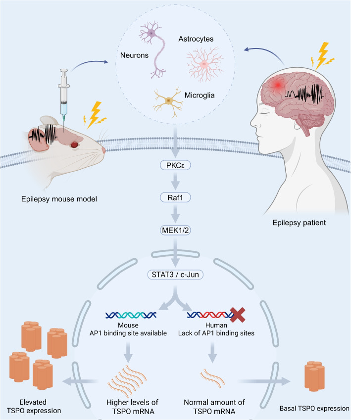
Translocator protein positron emission tomography (TSPO-PET) is a novel imaging modality that leverages the high expression of TSPO in activated microglia and other cells within seizure foci. It has been increasingly applied in the preoperative evaluation of drug-resistant epilepsy (DRE) to aid in the localization of these foci. With advances in tracer development, TSPO-PET has achieved higher signal-to-noise ratios and broader clinical utility. Clinical studies indicate that TSPO-PET yields significantly higher positive detection rates for seizure foci compared to magnetic resonance imaging and fluorodeoxyglucose positron emission tomography. This review summarizes recent progress in TSPO-PET radiotracer technology, its mechanism of action, and its clinical applications for managing DRE.
Keywords: Drug-resistant epilepsy; Neuroinflammation; Positron emission tomography; Seizure foci; Translocator protein.
© 2025. The Author(s).
Conflict of interest statement
Declarations. Ethics approval and consent to participate: Not applicable. Consent for publication: Not applicable. Competing interests: The authors declare that they have no competing interests.
Figures
Similar articles
-
Emerging translocator protein-positron emission tomographic imaging improves detection of focal cortical dysplasia.Epilepsia. 2025 Jul;66(7):2339-2352. doi: 10.1111/epi.18351. Epub 2025 Mar 8. Epilepsia. 2025. PMID: 40055993
-
Persistent neuroinflammation and cognitive impairment in a rat model of acute diisopropylfluorophosphate intoxication.J Neuroinflammation. 2016 Oct 12;13(1):267. doi: 10.1186/s12974-016-0744-y. J Neuroinflammation. 2016. PMID: 27733171 Free PMC article.
-
Preclinical characterization of [18F]D2-LW223: an improved metabolically stable PET tracer for imaging the translocator protein 18 kDa (TSPO) in neuroinflammatory rodent models and non-human primates.Acta Pharmacol Sin. 2025 Feb;46(2):393-403. doi: 10.1038/s41401-024-01375-9. Epub 2024 Aug 29. Acta Pharmacol Sin. 2025. PMID: 39210042
-
123I-MIBG scintigraphy and 18F-FDG-PET imaging for diagnosing neuroblastoma.Cochrane Database Syst Rev. 2015 Sep 29;2015(9):CD009263. doi: 10.1002/14651858.CD009263.pub2. Cochrane Database Syst Rev. 2015. PMID: 26417712 Free PMC article.
-
¹⁸F-FDG PET/CT: a review of diagnostic and prognostic features in multiple myeloma and related disorders.Clin Exp Med. 2015 Feb;15(1):1-18. doi: 10.1007/s10238-014-0308-3. Epub 2014 Sep 14. Clin Exp Med. 2015. PMID: 25218739
References
-
- Hong Z, Jiang YW. Clinical Guidelines. Epilepsy Volume: 2023 Revised Edition. Beijing: People’s Medical Publishing House; 2023. (In Chinese)
-
- Kalilani L, Sun X, Pelgrims B, Noack-Rink M, Villanueva V. The epidemiology of drug-resistant epilepsy: A systematic review and meta-analysis. Epilepsia. 2018;59(12):2179–93. - PubMed
-
- Chassoux F, Artiges E, Semah F, Laurent A, Landré E, Turak B, et al. 18F-FDG-PET patterns of surgical success and failure in mesial temporal lobe epilepsy. Neurology. 2017;88(11):1045–53. - PubMed
-
- Guo K, Wang J, Cui B, Wang Y, Hou Y, Zhao G, et al. [18F]FDG PET/MRI and magnetoencephalography may improve presurgical localization of temporal lobe epilepsy. Eur Radiol. 2022;32(5):3024–34. - PubMed
-
- Téllez-Zenteno JF, Hernández Ronquillo L, Moien-Afshari F, Wiebe S. Surgical outcomes in lesional and non-lesional epilepsy: a systematic review and meta-analysis. Epilepsy Res. 2010;89(2–3):310–8. - PubMed
Publication types
Grants and funding
LinkOut - more resources
Full Text Sources