. 2025 Aug 20;25(1):1342.
doi: 10.1186/s12903-025-06631-w.

The toxic effect of titanium dioxide nanoparticles on rat submandibular salivary glands and the protective role of vitamin E

Affiliations

The toxic effect of titanium dioxide nanoparticles on rat submandibular salivary glands and the protective role of vitamin E

Mariam Aboayana et al. BMC Oral Health. .

Abstract

Background: To assess the impact of titanium dioxide nanoparticles (TiO2NPs) on submandibular salivary glands and the role of vitamin E in preventing this cytotoxicity.

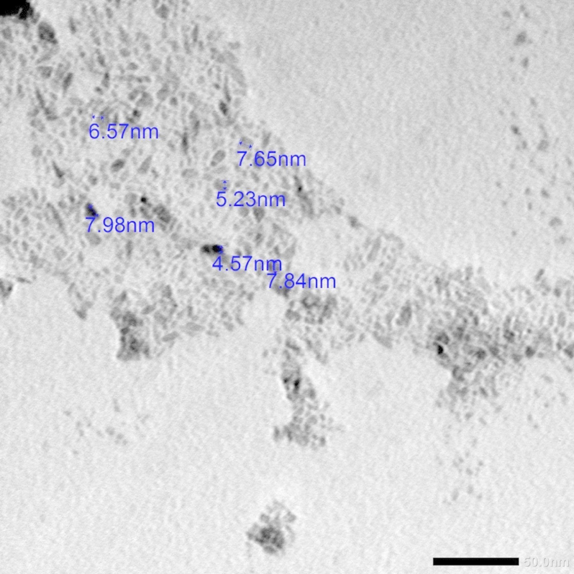
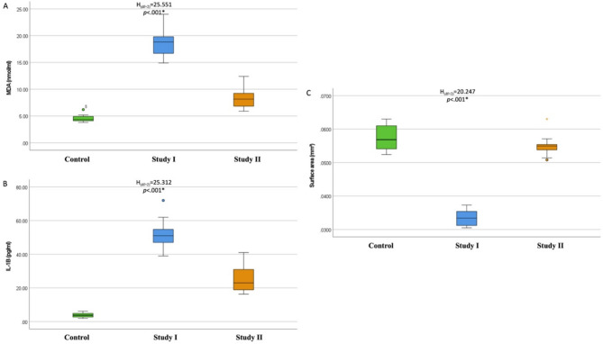
Methods: Thirty adult male albino rats were randomly divided into 3 groups: Negative control received olive oil for 3 weeks; Study I received olive oil for 1 week, then daily oral administration of 300 mg/kg TiO2NPs for 2 weeks; and Study II received 100 mg/kg vitamin E diluted in 100 ml olive oil daily as a prophylactic from day 1 for 3 weeks. On day 8, with vitamin E, they received 300 mg/kg TiO2NPs for 2 weeks by oral gavage. All samples were examined via hematoxylin & eosin (H&E), histomorphometry of serous acinar surface areas, transmission electron microscopy (TEM), and blood analysis of malondialdehyde (MDA) and interleukin (IL-1β) levels.

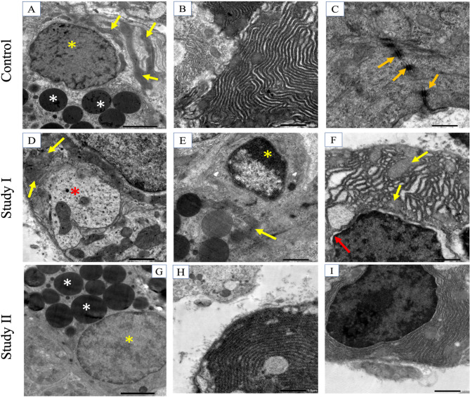
Results: Serum levels of both MDA and IL-1β were significantly greater in study I than in control and study II groups. Histologic examination revealed structural changes in serous acini and ducts of study I, with great preservation of the normal appearance of the acini and ducts in study II. Histomorphometry revealed a significant difference between control and study I, with no significant difference from that in study II. TEM revealed multiple ultrastructural changes in acinar cells and ducts of study I compared with those of study II, which maintained their normal features.

Conclusions: Vitamin E plays crucial antioxidant and anti-inflammatory roles in counteracting the cytotoxic effects of TiO2NPs by alleviating their deleterious impact on salivary glands.

Keywords: Nanoparticles; Nanotechnology; Submandibular glands; TiO2; Vitamin E.


Conflict of interest statement

Declarations. Competing interests: The authors declare no competing interests. Ethical approval: The Research Ethics Committee, Faculty of Dentistry, Alexandria University, approved the current study IRB: 0678-5/2023. The current experiment complies with the ARRIVE guidelines, EU Directive 2010/63/EU for animal experiments and the National Research Council’s Guide for the Care and Use of Laboratory Animals [56]. Informed consent: Not applicable. Clinical trial number: not applicable.

Figures

Fig. 1
Fig. 1
Transmission electron micrograph (TEM) showing TiO2NPs less than 100 nm in size (TEM ×100,000. Scale bar 50 nm)
Fig. 2
Fig. 2
Box and whisker graph. The thick line in the middle of the box represents the median, the box represents the interquartile range (from the 25th to 75th percentiles), and the whiskers represent the minimum and maximum values. 2A: MDA (nmol/ml) serum levels, 2B: interleukin 1-beta (IL-1β, pg/ml) serum levels, and 2 C: mean surface area (SA) of the serous acini (mm2)
Fig. 3
Fig. 3
Light micrographs (LM) of a rat submandibular gland, H&E stain, control [3 A, 3B, 3 C, 3D], (3 A, 3B) serous acini (black asterisk), striated ducts (red asterisk), and granular convoluted ducts (yellow arrow) ×400. (3 C) Excretory duct (green asterisk) ×100. (3D) Higher magnification of the excretory duct ×400. Study I [3E, 3 F, 3G, 3 H], (3E) Nuclear pleomorphism and abnormal appearance of serous acini (black asterisk) striated ducts (red asterisk) x400. (3 F) Abnormally granular convoluted ducts (yellow arrow). Nuclear pleomorphism of serous acini (black asterisk) and striated ducts (red asterisk) x400. (3G) Degeneration of fibrous tissue (green asterisk) ×100. Note the large interacinar spaces (orange asterisk). (3 H) Higher magnification image of the excretory duct ×400. Study II (3I, 3 J, 3 K, 3 L). (3I, 3 J) Serous acini (black acini), striated (red asterisk), and granular convoluted ducts (yellow arrow) x400. (3 K) Excretory duct (green asterisk) ×100. (3 L) Higher magnification of the excretory duct x400
Fig. 4
Fig. 4
TEM image showing the salivary acini of the control (4 A, 4000×) active nucleus of serous acini (yellow asterisk) and uniform secretory globules (white asterisk). mitochondria present perinuclearly (yellow arrows). (4B, x6000) rER. (4 C, ×12000) Cellular junctions (orange arrows). Study I (4D, ×6000) serous acinar cells revealing cytoplasmic degeneration (red asterisk). Loss of mitochondrial cristae (yellow arrows). (4E, x6000) Chromatin condensation (yellow asterisk). Loss of mitochondrial striations (yellow arrow). (4 F, ×6000) Abnormal appearance of the rER surrounding the nucleus of the acinar cell along with loss of the striations of mitochondria (yellow arrows) and blebbing of the nuclear membrane (red arrow). Study II (4G, x4000) revealed the active nucleus of the acinar cell (yellow asterisk) and secretory granules (white asterisk). (4 H, 4I, ×6000) Normal appearance of the rER
Fig. 5
Fig. 5
TEM image showing salivary ducts, control, (5 A, x800) intercalated duct, (5B, x4000) active nucleus (yellow asterisk) of the intercalated duct, (5 C, x4000) striated duct showing distinct basal foldings with regularly organized mitochondria (yellow arrows). Study I, (5D, ×6000) Intercalated duct with pyknotic apoptotic nucleus (yellow asterisk), tertiary lysosome (square area), and abnormal intercellular attachment (orange arrows). (5E, x4000) Striated duct showing loss of basal striations, disorganized apoptotic mitochondria (square area), and irregularly shaped fused mitochondria (yellow arrow). (5 F, ×6000) Pyknotic nucleus (asterisk) of the striated duct. Study II: (5G, x6000) Active nuclei of the intercalated duct (yellow asterisk), separation between the nucleus and cytoplasm (red arrow). (5 H, ×12000) Higher magnification of the previous image showing intact cellular membranes and desmosomes (orange arrows). (5I, x6000) Striated duct with evident basal striations. Well-defined cristae of the mitochondria (yellow arrows)

References

    1. Abdel Aal SM, Ahmed SM, Abdelrahman SA, Abdelrahman AA, Samy W. Duration-dependent effects induced by titanium dioxide nanoparticles on pancreas of adult male albino rats (histological and biochemical study). Ultrastruct Pathol. 2020;44:342–58. 10.1080/01913123.2020.1786203. - PubMed
    1. Chen Z, Han S, Zheng P, Zhou D, Zhou S, Jia G. Effect of oral exposure to titanium dioxide nanoparticles on lipid metabolism in Sprague–Dawley rats. Nanoscale. 2020;12:5973–86. 10.1039/C9NR10947A. - PubMed
    1. Musial J, Krakowiak R, Mlynarczyk DT, Goslinski T, Stanisz BJ. Titanium dioxide nanoparticles in food and personal care products—what do we know about their safety? Nanomaterials. 2020;10(6):1110. 10.3390/nano10061110. - PMC - PubMed
    1. Stefaniak A, Hoover M, Nanotechnology N. Occupational Exposure to Titanium Dioxide [Internet]. Available from: www.cdc.gov/niosh
    1. Bischoff NS, de Kok TM, Sijm DTHM, van Breda SG, Briedé JJ, Castenmiller JJM, et al. Review possible adverse effects of food additive e171 (Titanium dioxide) related to particle specific human toxicity, including the immune system. Int J Mol Sci. 2021;22(1):207. 10.3390/ijms22010207. - PMC - PubMed

LinkOut - more resources