Review
. 2025 Sep 3;73(35):21701-21727.
doi: 10.1021/acs.jafc.5c03663. Epub 2025 Aug 21.

Pheromone-Binding Proteins in Pest Control: From Molecular Insights to Real-World Applications

Affiliations
Review

Pheromone-Binding Proteins in Pest Control: From Molecular Insights to Real-World Applications

Ishani Ray et al. J Agric Food Chem. .

Abstract

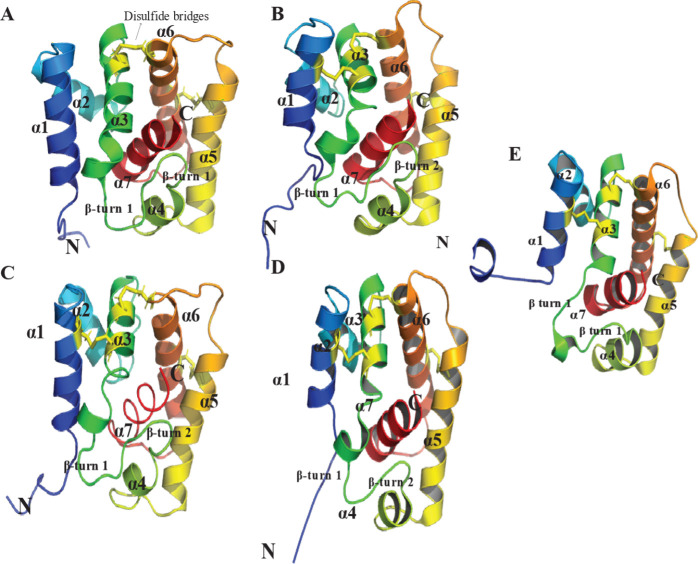
Insects utilize sophisticated olfactory systems to detect chemical cues critical for behaviors such as mating, host selection, and predator avoidance. In lepidopteran moths, sex pheromone communication offers a well-established model in which males detect female-emitted signals over long distances. Central to this process are pheromone-binding proteins (PBPs), which solubilize and transport hydrophobic pheromones through the sensillar lymph to olfactory receptors, enabling precise signal detection. Recent advances in molecular biology, structural biochemistry, and gene-editing technologies such as CRISPR/Cas9 have uncovered nuanced mechanisms underlying PBP function, including ligand-binding specificity, pH-dependent conformational dynamics, and molecular interactions. These discoveries have broad implications, extending beyond chemosensory biology to applications in reverse chemical ecology, biosensing, and environmentally conscious pest control. This review synthesizes insights from in vitro, in silico, and in vivo studies, highlighting the structural and functional diversity of PBPs across species and emphasizing their translational utility as molecular targets for sustainable agriculture and biodiversity conservation.

Keywords: integrated pest management (IPM); lepidopteran sex pheromones; ligand binding; odorant-binding proteins (OBPs); olfaction in insects; olfactory receptors (ORs); pest control strategies; pheromone-binding proteins (PBPs); signal transduction.


Figures

1
1
Moth olfactory sensillum structure. The image illustrates the moth’s antenna, which houses specialized olfactory sensilla responsible for detecting pheromones. A magnified cross-section of a sensillum shows its structural components, including cuticle pores, lymph, dendrites of olfactory sensory neurons, and olfactory receptors. Created in BioRender in collaboration with Smita Mohanty. Ray, I. (2025) https://BioRender.com/05ei6au.
2
2
Schematic representation of sensillar environment, pheromone transport and odorant detection pathways. Key components include olfactory receptors (ORs), coreceptors (Orco), sensory neuron membrane proteins (SNMP), odorant sensory neurons (OSNs), pheromone-degrading enzymes (PDEs) and pheromone-binding proteins (PBPs). After transport through the sensillar lymph, the pheromone–PBP complex binds to the OrX/Y–Orco receptor complex, activating ionotropic (A) or metabotropic (B) signaling pathways. The ionotropic (A) pathway directly allows calcium influx; the metabotropic pathway (A) involves G-protein signaling, activating phospholipase C (PLC), which hydrolyzes PIP2 into DAG and IP3, leading to further calcium release. However, the role of the metabotropic pathway in insect olfactory transduction remains debated, with current evidence suggesting it plays a relatively minor role. The schematic reflects both proposed mechanisms. Created in BioRender in collaboration with Smita Mohanty. Ray, I. (2025) https://BioRender.com/8g34e6m.
3
3
Representative structural organization of the pheromone-binding pocket in moth PBPs. The hydrophobic pocket is formed by four core helices (α1, α4, α5, and α6) converging at the narrow end, while helix α3 caps the opposite end.
4
4
Schematic representation of pheromone binding and release mechanism by pheromone-binding proteins (PBPs). Representative pH-Induced Conformational Changes shown within the schema using ApolPBP1 structures: the open conformation at pH 4.5 (PDB: 2JPO) facilitates ligand release, while the closed conformation at pH 6.5 (PDB: 1QWV) enables ligand binding. The C-terminus in red in both states. Created in BioRender in collaboration with Smita Mohanty. Ray, I. (2025) https://BioRender.com/65itn35.
5
5
Structural Comparison of Moth PBP A-Form Conformations. This figure illustrates the A-form structures of moth PBPs, labeling helices α1-α7 and β-turn motifs. Conserved disulfide linkages are shown as sticks in yellow. (A) BmorPBP1 (PDB ID: 1GM0), averaged NMR structure at pH 4.5, (B) BmorPBP1 (PDB ID: 2FJY), X-ray structure at 2.30 Å resolution, delipidated, pH 7.5, (C) ApolPBP1 (PDB ID: 2JPO), averaged NMR structure at pH 4.5, (D) AtraPBP1 (PDB ID: 2KPH), lowest energy NMR structure at pH 4.5, (E) LdisPBP1 (PDB ID: 6UM9), averaged NMR structure at pH 4.5. Structural visualization was performed using PyMOL.
6
6
NMR Structures of Moth PBPs in the B-Form: (A) BmorPBP1 (PDB ID: 1LS8, pH 6.5), (B) ApolPBP1 (PDB ID: 1QWV, pH 6.3). Structural visualization was performed using PyMOL.
7
7
Representative comparison of histidine gates between open and closed conformations at different pH conditions. The protein backbone is in red, H residues in purple and the C-terminus in light blue. (A) NMR structure of ApolPBP1 (PDB ID: 1QWV) at pH 6.3, unstructured C-terminus and 6.40 Å between H70 and H95, (B) NMR structure of ApolPBP1 (PDB ID: 2JPO) at pH 4.5, C-terminal α-helix and 12.62 Å between H70 and H95. Adapted with permission under a Creative Commons CC-BY 4.0 (https://creativecommons.org/licenses/by/4.0/) from [128]. Colors and labels have been modified for clarity. Copyright 2009 ASBMB, currently Elsevier. Structural visualization was performed using PyMOL.
8
8
C-Terminal Helices of the A-Form PBPs with acidic residues in sticks: (A) BmorPBP1 (PDB ID: 1GM0) with interactions within 4 Å of acidic residues highlighted, (B) BmorPBP1 (PDB ID: 2FJY), (C) ApolPBP1 (PDB ID: 2JPO), (D) AtraPBP1 (PDB ID: 2KPH), (E) LdisPBP1 (PDB ID: 6UM9). Structural visualization was performed using PyMOL.
9
9
Ligand-Protein Interactions in Moth PBP-Ligand Complexes. The top figures present crystallographic structures of moth PBPs bound to their respective ligands with helices color-coded. The bottom figures highlight the key protein residues (stick models superimposed on surface depiction of protein) involved in ligand (stick models) binding. (A) BmorPBP1 complexed with bombykol (PDB ID: 1DQE, 1.80 Å resolution) at pH 8.0, (B) AtraPBP1 complexed with Z11, Z13–16: Ald (PDB ID: 4INW, 1.14 Å resolution) at pH 6.5, (C) AtraPBP1 complexed with Z11, Z13–16: OH (PDB ID: 4INX, 1.85 Å resolution) at pH 6.5, (D) Binding site residues of BmorPBP1 with bombykol, (E) Binding site residues of AtraPBP1 with the Z11,Z13–16:Ald, (F) Binding site residues of AtraPBP1 with Z11,Z13–16:OH. Structural visualization was performed using PyMOL.
10
10
Averaged NMR structures of OfurPBP2 at pH 6.5 (PDB ID: 7UO6). Helices are colored from blue (N-terminus) to red (C-terminus): (A) with a well-structured C-terminal helix (α7), (B) Arg70-His88 gate shown as stick representations in violet. Structural visualization was performed using PyMOL.

Similar articles

References

    1. Hildebrand J. G., Shepherd G. M.. Mechanisms of olfactory discrimination: Converging evidence for common principles across phyla. Annu. Rev. Neurosci. 1997;20:595–631. doi: 10.1146/annurev.neuro.20.1.595. - DOI - PubMed
    1. Cande J., Prud’homme B., Gompel N.. Smells like evolution: the role of chemoreceptor evolution in behavioral change. Curr. Opin. Neurobiol. 2013;23:152–158. doi: 10.1016/j.conb.2012.07.008. - DOI - PubMed
    1. Regnier F. E., Law J. H.. Insect Pheromones. J. Lipid Res. 1968;9(5):541. doi: 10.1016/S0022-2275(20)42699-9. - DOI - PubMed
    1. Greenfield M. D.. Moth Sex-Pheromones - an Evolutionary Perspective. Fla Entomol. 1981;64(1):4–17. doi: 10.2307/3494597. - DOI
    1. Honson N., Gong Y., Plettner E.. Structure and function of insect odorant and pheromone-binding proteins (OBPs and PBPs) and chemosensory-specific proteins (CSPs) Recent Advances in Phytochemistry. 2005;39(05):227–268. doi: 10.1016/S0079-9920(05)80010-3. - DOI

MeSH terms