

Shear wave elastography reveals elevated infrapatellar fat pad stiffness in patients with early osteoarthritis symptoms after ACL reconstruction
- PMID: 40836995
- PMCID: PMC12362417
- DOI: 10.1016/j.ostima.2025.100267
Shear wave elastography reveals elevated infrapatellar fat pad stiffness in patients with early osteoarthritis symptoms after ACL reconstruction
Abstract
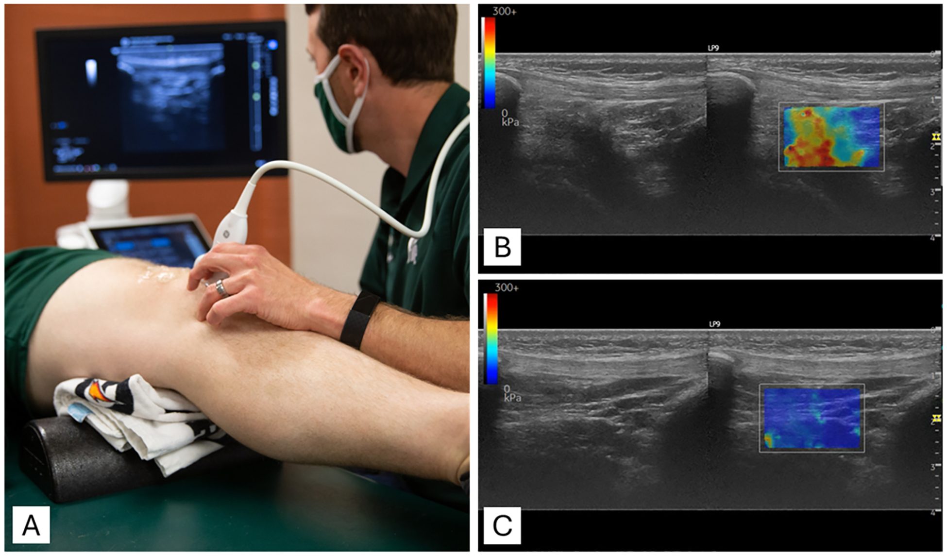
Objective: The infrapatellar fat pad (IPFP) plays an important role in knee biomechanics and inflammation, particularly following anterior cruciate ligament reconstruction (ACLR). This study investigated whether IPFP stiffness, measured with shear wave elastography, is associated with early symptoms of osteoarthritis (OA) in individuals within one year after ACLR.
Design: In this cross-sectional study, 24 participants underwent bilateral IPFP stiffness assessments using shear wave elastography. Participants were positioned supine with 20° knee flexion. The stiffness limb symmetry index (LSI) was calculated to normalize stiffness between the ACLR and contralateral limbs. Early OA symptoms were defined as scores ≤85 % on at least two of four subscales of the Knee Injury and Osteoarthritis Outcome Score (KOOS). Independent t-tests were used to evaluate group differences in IPFP stiffness LSI, and receiver operating characteristic curve analysis determined the optimal LSI threshold for discriminating between groups.
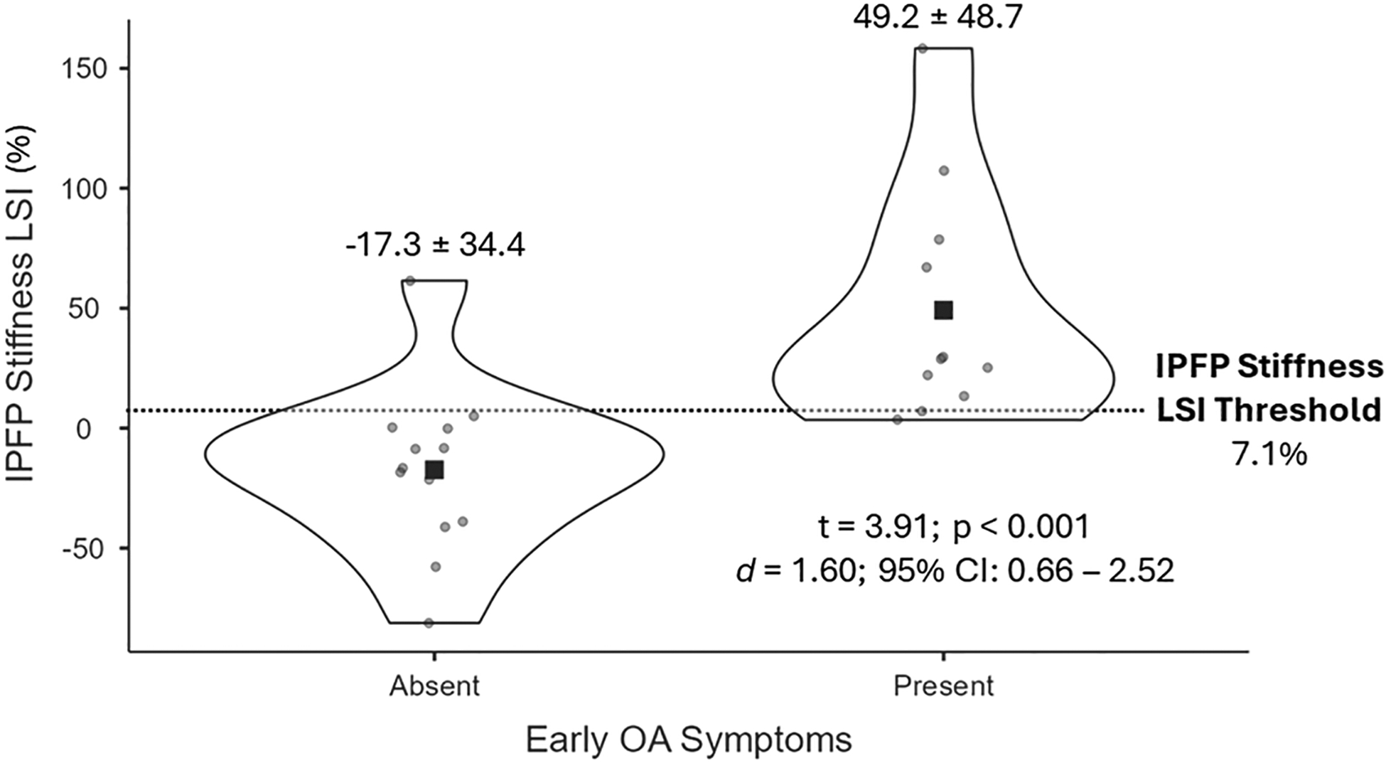
Results: Eleven participants (46 %) showed early OA symptoms. Participants with early OA symptoms exhibited a significantly higher IPFP stiffness LSI compared to those without symptoms (49.2 ± 48.7 % vs. -17.3 ± 34.4 %, p < 0.001). An optimal stiffness LSI threshold of 7.1 % was identified, achieving 90.9 % sensitivity, 92.3 % specificity, and an area under the curve of 0.94.
Conclusions: Shear wave elastography shows potential as a non-invasive tool for detecting early IPFP stiffness changes associated with OA symptoms post-ACLR. These findings suggest that IPFP stiffness may be an early marker for OA risk, warranting further longitudinal studies to evaluate its progression and to further examine the clinical utility of shear wave elastography.
Keywords: ACLR; Elastography; IPFP; Rehabilitation; Ultrasonography.
Conflict of interest statement
Conflict of interest None of the authors has any other financial interests that could create a potential conflict of interest or the appearance of a conflict of interest with regard to this work. Declaration of competing interest The authors declare that they have no known competing financial interests or personal relationships that could have appeared to influence the work reported in this paper.
Figures

Similar articles
-
Failure to Achieve the Patient Acceptable Symptom State 2 Years After Anterior Cruciate Ligament Reconstruction Reflects Poor Knee Loading Patterns.Am J Sports Med. 2025 Jul;53(9):2136-2144. doi: 10.1177/03635465251349105. Epub 2025 Jun 26. Am J Sports Med. 2025. PMID: 40566928
-
A novel infrapatellar fat pad preservation technique in anterior cruciate ligament reconstruction reduces postoperative pain and cartilage damage: a retrospective study.J Orthop Surg Res. 2025 Aug 5;20(1):728. doi: 10.1186/s13018-025-06162-8. J Orthop Surg Res. 2025. PMID: 40765021 Free PMC article.
-
Surgical versus conservative interventions for treating anterior cruciate ligament injuries.Cochrane Database Syst Rev. 2016 Apr 3;4(4):CD011166. doi: 10.1002/14651858.CD011166.pub2. Cochrane Database Syst Rev. 2016. PMID: 27039329 Free PMC article.
-
Using 3D MRI Bone Shape to Predict Pre-Osteoarthritis of the Knee 2 Years After Anterior Cruciate Ligament Reconstruction.Am J Sports Med. 2023 Dec;51(14):3677-3686. doi: 10.1177/03635465231207615. Epub 2023 Nov 7. Am J Sports Med. 2023. PMID: 37936374 Free PMC article.
-
Braces and orthoses for treating osteoarthritis of the knee.Cochrane Database Syst Rev. 2015 Mar 16;2015(3):CD004020. doi: 10.1002/14651858.CD004020.pub3. Cochrane Database Syst Rev. 2015. PMID: 25773267 Free PMC article.
References
-
- Zhou S, et al. , Source and hub of inflammation: the infrapatellar fat pad and its interactions with articular tissues during knee osteoarthritis, J. Orthop. Res 40 (7) (2022) 1492–1504. - PubMed
-
- Han W, et al. , Signal intensity alteration in the infrapatellar fat pad at baseline for the prediction of knee symptoms and structure in older adults: a cohort study, Ann. Rheum. Dis 75 (10) (2016) 1783–1788. - PubMed
Grants and funding
LinkOut - more resources
Full Text Sources