. 2025 Jul 30;28(9):113251.
doi: 10.1016/j.isci.2025.113251. eCollection 2025 Sep 19.

Functional characterization of the Csm1-like protein TITAN 9 in Arabidopsis thaliana

Affiliations

Functional characterization of the Csm1-like protein TITAN 9 in Arabidopsis thaliana

Zhijian Zhang et al. iScience. .

Abstract

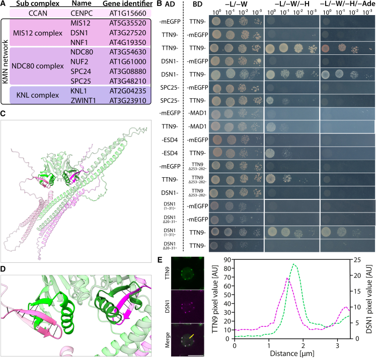
Kinetochores are essential for chromosome segregation in eukaryotes. An important component of kinetochores in opisthokonta is Csm1. However, its function appears to be diversified and, while Csm1 in budding yeast is a component of the monopolin complex mediating mono-orientation of sister kinetochores during meiosis I, the fission yeast homolog Pcs1 prevents merotelic spindle microtubule attachments during mitosis and meiosis II. Here, we have characterized TITAN9 (TTN9), a distantly related Csm1-like protein in the flowering plant Arabidopsis. TTN9 accumulates in mitotic and meiotic tissue and localizes to centromeres throughout the cell cycle. By analyzing proteome-wide TTN9 associated proteins, we identified a substantial subset of the Arabidopsis kinetochore proteome, including DSN1, mirroring known Csm1 interactions in yeast. While homozygous ttn9 mutants are not viable, a meiosis-specific knock-down of TTN9 causes chromosome segregation defects and split centromeres during meiosis I. These findings suggest that Csm1-like proteins contribute to conserved kinetochore functions across eukaryotes.

Keywords: plant Biology; plant bioinformatics; plant development; plant evolution; plant genetics; plant physiology; plant systematics.


Conflict of interest statement

The authors declare no competing interests.

Figures

None
Graphical abstract
Figure 1
Figure 1
Subcellular localization of TTN9 during mitosis and interphase in root cells Scale bars: 10 μm. (A) TTN9-GFP (green) accumulates in all tissue layers of the root. Microtubules were visualized by RFP-TUA5 (magenta). (B) Localization of TTN9-GFP (green) and RFP-TUA5 (magenta) during mitosis. (C) Localization of TTN9-GFP (green) and RFP-CENH3 (magenta) during interphase. The white arrowhead indicates TTN9-GFP localization at the nuclear periphery. (D) TTN9-GFP (green) signal largely overlaps with RFP-CENH3 signal; position of the line scan is indicated by a yellow line in C.
Figure 2
Figure 2
Subcellular localization of TTN9 during meiosis Localization pattern of TTN9-GFP (green) in male meiocytes. Microtubule structures were visualized by RFP-TUA5 (magenta). Scale bars: 10 μm.
Figure 3
Figure 3
TTN9 is part of the KMN network (A) Kinetochore components that were precipitated with TTN9 as a bait. (B) TTN9 interacts with itself, SPC25, MAD1, DSN1, and ESD4 in Y2H assays. AD, activating domain; BD, DNA-binding domain. The position of the AD or the BD tag is indicated by a dash. Yeast cells were diluted and spotted on different dropout media as indicated on top. Growth on −L/−W/−H and −L/−W/−H/−Ade indicates interaction. Negative interaction assays between TTN9 and other kinetochore components are shown in Figure S2. (C) AlphaFold-Multimer model of 2×TTN9 (green) and 2×DSN1 (magenta); regions deleted in the Y2H deletion constructs DSN1Δ20-31 and TTN9Δ253-282 used in B are shown in more saturated color. (D) Magnification of the predicted TTN9–DSN1 interaction regions, H-bonds emanating from DSN1 amino acids 20–31 are shown as dashed lines. (E) TTN9 (green) partially co-localizes with DSN1 (magenta) in somatic interphase cells; the position of the line scan is indicated by a yellow line. Scale bar: 10 μm.
Figure 4
Figure 4
TTN9 is important for faithful chromosome segregation in meiosis Scale bars: 10 μm. (A) Localization pattern of BMF1-GFP (green) and RFP-TUA5 (magenta) in WT and amiTTN9-1 meiocytes. White arrowheads highlight mis-congressed (metaphase I) or lagging (anaphase I) chromosomes. (B) Quantification of back and forth moving (“dancing”) chromosomes. Bars represent the percentage of cells with “dancing” chromosomes. Significant differences, as determined by Fisher’s exact test (p < 0.001, Bonferroni-corrected), are indicated by differing letters over the bars. (C) Quantification of NEB-to-AO duration in WT and amiTTN9-1 meiocytes. For tNEB–C (duration from NEB to full congression) and tC–AO (duration from full congression to AO), only movies where chromosomes eventually congressed were analyzed. Data are represented as median ± interquartile range; whiskers indicate the full range, excluding outliers. Significant differences, as determined by Student’s t test (p < 0.05), are indicated by differing letters over the bars. (D) Unbalanced pools of chromosomes after metaphase I observed in FISH-stained meiocytes (white: DAPI; green: centromeres). Bars represent the percentage of cells with unbalanced pools. (E) Metaphase I in WT compared to amiTTN9-1 and amiTTN9-2 meiocytes. In amiTTN9-1 and amiTTN9-2 plants, extra centromere signals on chromosomes indicate split sister centromeres. Chromosomes encircled by a dashed blue line show examples of WT-like fused sister centromeres. Examples of chromosomes with split sister centromeres are highlighted by ovals with dashed orange lines, respectively. Split centromere signals are indicated by arrowheads. (F) Occurrence of split sister centromeres in WT, amiTTN9-1, and amiTTN9-2. Bars represent the percentage of cells with split sister centromeres. Significant differences were determined using Fisher’s exact test with Holm correction for multiple comparisons. Asterisks indicate significant differences compared to WT (∗p < 0.05; ∗∗p < 0.01; ∗∗∗p < 0.001).
Figure 5
Figure 5
Model of TTN9 action (A) Schematic representation of the outer kinetochore architecture in Arabidopsis thaliana, based on known KMN network interactions. Solid black lines indicate binary interactions found in this study. Dashed lines indicate weak interactions in our Y2H assay that are not well supported by AlphaFold. NSL1 was the only component not found in the interactome of TTN9 in this study. The link between the KMN network and the CCAN remains unresolved. (B) Different functions of TTN9 in somatic tissue. During mitosis and/or mitotic interphase, TTN9 plays an essential but so far unknown role. KT, kinetochore associated function; NE, nuclear envelope associated function. (C) Different functions of TTN9 in meiosis. TTN9’s NE-associated localization might indicate a function in preventing illegitimate recombination, albeit evidence is still lacking for plants. During meiosis I, TTN9 might clamp MT binding sites of the same and/or sister kinetochores, enforcing monopolar attachments of sister kinetochores and/or preventing merotelic attachments.

References

    1. Chelysheva L., Diallo S., Vezon D., Gendrot G., Vrielynck N., Belcram K., Rocques N., Márquez-Lema A., Bhatt A.M., Horlow C., et al. AtREC8 and AtSCC3 are essential to the monopolar orientation of the kinetochores during meiosis. J. Cell Sci. 2005;118:4621–4632. - PubMed
    1. Yokobayashi S., Watanabe Y. The kinetochore protein Moa1 enables cohesion-mediated monopolar attachment at meiosis I. Cell. 2005;123:803–817. - PubMed
    1. Watanabe Y., Nurse P. Cohesin Rec8 is required for reductional chromosome segregation at meiosis. Nature. 1999;400:461–464. - PubMed
    1. Ogushi S., Rattani A., Godwin J., Metson J., Schermelleh L., Nasmyth K. Loss of sister kinetochore co-orientation and peri-centromeric cohesin protection after meiosis I depends on cleavage of centromeric REC8. Dev. Cell. 2021;56:3100–3114.e4. - PMC - PubMed
    1. Sakuno T., Tada K., Watanabe Y. Kinetochore geometry defined by cohesion within the centromere. Nature. 2009;458:852–858. - PubMed

LinkOut - more resources