. 2025 Aug 15;16(8):108101.
doi: 10.4239/wjd.v16.i8.108101.

Hyperglycemia-induced overexpression of CREB3 L3 promotes the epithelial-to-mesenchymal transition in bladder urothelial cells in diabetes mellitus

Affiliations

Hyperglycemia-induced overexpression of CREB3 L3 promotes the epithelial-to-mesenchymal transition in bladder urothelial cells in diabetes mellitus

Qing-Guo Wu et al. World J Diabetes. .

Abstract

Background: Diabetic cystopathy (DCP) is a complication affecting the lives of people with diabetes. However, the pathogenesis of DCP is not well known.

Aim: To investigate the potential mechanisms by which cAMP-responsive element-binding protein 3 like 3 (CREB3 L3) promotes the occurrence and development of DCP.

Methods: High-throughput sequencing was used to analyze differentially expressed genes (DEGs) in bladder urothelium from patients with DCP and healthy controls. Gene enrichment analysis was conducted to assess the biological functions of DEG. Small interfering RNA technology was performed to silence the CREB3 L3 gene in both in vitro and in vivo experiments. Morphological changes in bladder urothelium from a DCP rat model were observed. Immunofluorescence and western blotting assay were performed to determine associated protein expression.

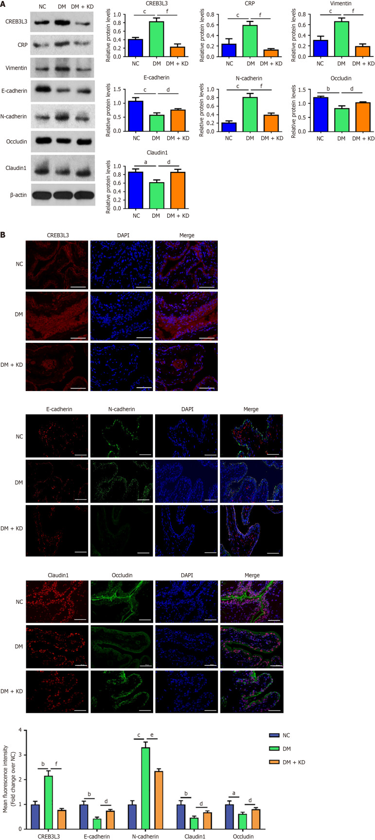
Results: We identified significant DEGs through high-throughput sequencing. These genes were primarily enriched in inflammatory activation, epithelial-mesenchymal transition (EMT) and tight junction organization. Upregulated expression of both CREB3 L3 and C-reactive protein (CRP) in bladder urothelium from patients with DCP was accompanied by upregulated EMT markers including N-cadherin and vimentin proteins, but downregulated E-cadherin. Silencing CREB3 L3 attenuated the protein expression of CRP and EMT in SV-HUC-1 urothelial cells under hyperglycemic conditions and in the diabetes mellitus rat model at 4, 8, and 12 weeks. CREB3 L3 knockdown also reversed downregulation of the tight junction proteins occludin and claudin 1.

Conclusion: Hyperglycemia induces the upregulation of CREB3 L3 expression, thereby promoting the EMT and impairing tight junctions in bladder urothelial cells. Targeting CREB3 L3 in bladder urothelial cells is likely to be a key approach in preventing and treating DCP.

Keywords: Diabetic cystopathy; Epithelial-mesenchymal transition; Hyperglycemia.


Conflict of interest statement

Conflict-of-interest statement: The authors declare that they have no known competing financial interests or personal relationships that could have appeared to influence the work reported in this paper.

Figures

Figure 1
Figure 1
Differentially expressed cAMP-responsive element-binding protein 3 Like 3, C-reactive protein, and proteins related to the epithelial-mesenchymal transition and tight junctions in the bladder urothelial tissues of patients with diabetic cystopathy. Second-generation sequencing was conducted to find genes that are differentially expressed in bladder urothelial tissues between the diabetic cystopathy (DCP) (n = 3) and healthy control (HC) group (n = 3). A: Heatmap of differentially expressed genes (DEGs); B: Volcanic map of DEGs; C: Gene Ontology analysis of the DEGs, including biological process, cellular component, and molecular function; D: Kyoto Encyclopedia of Genes and Genomes analysis of DEGs; E: To identify the results from the second-generation sequencing, we performed western blotting to detect the protein expression of bladder urothelium from the DCP group (n = 6) and HC group (n = 6). aP < 0.01 vs HC group, bP < 0.01 vs HC group. CRP: C-reactive protein.
Figure 2
Figure 2
High glucose induces cAMP-responsive element-binding protein 3 Like 3 expression and the epithelial-mesenchymal transition in SV-HUC-1 cells. A: Cell Counting Kit-8 assay was employed to assess the viability of SV-HUC-1 cells following treatment with varying concentrations of glucose; B: Relative mRNA expression of cAMP-responsive element-binding protein 3 Like 3 (CREB3 L3) in SV-HUC-1 cells after treating with different levels of glucose, as detected by quantitative PCR; C: Relative mRNA expression of CREB3 L3 in SV-HUC-1 cells with or without silencing of CREB3 L; D: SV-HUC-1 cells were treated with 5 mmol/L and 15 mmol/L glucose for 72 hours with and without CREB3 L3 knockdown (KD). Protein expression of CREB3 L3, C-reactive protein (CRP), vimentin, N-cadherin, E-cadherin, and occludin in SV-HUC-1 cells in the control (Con), high glucose (HG), HG + small interfering RNA negative control (si-NC), and HG + KD group. Each group contained three biological replicates. aP < 0.05 vs 5 mmol/L group, bP < 0.01 vs 5 mmol/L group; cP < 0.001 vs 5 mmol/L group, dP < 0.05 vs si-NC group, eP < 0.01 vs si-NC group, fP < 0.001 vs si-NC group, gP < 0.01 vs Con group, hP < 0.001 vs Con group, iP < 0.01 vs HG + si-NC group, jP < 0.001 vs HG + si-NC group.
Figure 3
Figure 3
Immunofluorescence of cAMP-responsive element-binding protein 3 Like 3, C-reactive protein, E-cadherin, and occludin in urothelial cells under high glucose condition. SV-HUC-1 cells were treated with 5 mmol/L and 15 mmol/L glucose for 72 hours with and without cAMP-responsive element-binding protein 3 Like 3 (CREB3 L3) knockdown (KD). Immunofluorescence assay was conducted to detect the expression levels of CREB3 L3, C-reactive protein (CRP), E-cadherin, and occludin protein in SV-HUC-1 cells in the control (Con), high glucose (HG), HG + small interfering RNA negative control (si-NC), and HG + KD groups. Scale bar indicates 50 μm, and each group contained three replicates. aP < 0.01 vs Con group, bP < 0.001 vs Con group, cP < 0.05 vs HG + si-NC group, dP < 0.01 vs HG + si-NC group, eP < 0.001 vs HG + si-NC group.
Figure 4
Figure 4
Establishment of diabetic cystopathy models of rats for 4 weeks. A and B: This study established a diabetic cystopathy model in rats by feeding a high-fat and high-sugar diet and intraperitoneally injecting 1% streptozotocin; C-F: At the end of the 4-week period, the body weight, blood glucose, bladder weight and diameter of the rats were measured, and each group contained five replicates; G: The bladder tissue was retained for hematoxylin and eosin staining, and each group contained three replicates. aP < 0.01 vs negative control (NC) group, bP < 0.05 vs diabetes mellitus (DM) group.
Figure 5
Figure 5
cAMP-responsive element-binding protein 3 Like 3 upregulates C-reactive protein expression, induces the epithelial-mesenchymal transition, and impairs cellular tight junctions in the diabetic cystopathy rat model. A: Western blot analysis of cAMP-responsive element-binding protein 3 Like 3 (CREB3 L3), C-reactive protein (CRP), vimentin, N-cadherin, E-cadherin, claudin 1, and occludin in the bladder urothelial tissues from each group; B: Immunofluorescence analysis of the expression of CREB3 L3, N-cadherin, E-cadherin, claudin 1, and occludin proteins in the bladder urothelial tissues from each group. Each group contained three biological replicates. Scale bar indicates 50 μm. aP < 0.05 vs negative control (NC) group, bP < 0.01 vs NC group, cP < 0.001 vs NC group; dP < 0.05 vs diabetes mellitus (DM) group, eP < 0.01 vs DM group, fP < 0.001 vs DM group.
Figure 6
Figure 6
Establishment of diabetic cystopathy rat models for 8 weeks. A: At the end of the 8 weeks, the body weight, blood glucose and bladder weight from rats were measured; B: Hematoxylin and eosin staining of bladder tissues of the rats at the end of the 8 weeks, and each group contained three replicates; C: Western blotting detected the expression of cAMP-responsive element-binding protein 3 Like 3 (CREB3 L3), C-reactive protein (CRP), vimentin, N-cadherin, E-cadherin, claudin 1, and occludin in the bladder urothelial tissues of the rats at the end of 8 weeks. Each group contained three replicates. aP < 0.05 vs negative control (NC) group, bP < 0.01 vs NC group, cP < 0.001 vs NC group; dP < 0.05 vs diabetes mellitus (DM) group, eP < 0.01 vs DM group, fP < 0.001 vs DM group.
Figure 7
Figure 7
cAMP-responsive element-binding protein 3 Like 3 continuously induces the epithelial-mesenchymal transition and impairs cellular tight junctions with the progression of diabetic cystopathy. At the end of 8 weeks, immunofluorescence staining of cAMP-responsive element-binding protein 3 Like 3 (CREB3 L3), N-cadherin, E-cadherin, claudin 1, and occludin proteins in the bladder urothelial tissues from each group. Each group contained three replicates. Scale bar indicates 50 μm. aP < 0.05 vs negative control (NC) group, bP < 0.01 vs NC group, cP < 0.001 vs NC group, dP < 0.0001 vs NC group; eP < 0.05 vs diabetes mellitus (DM) group, fP < 0.01 vs DM group, gP < 0.001 vs DM group.
Figure 8
Figure 8
Establishment of diabetic cystopathy models of rats for 12 weeks. A: At the end of the 12 weeks, the body weight, blood glucose and bladder weight from rats were measured; B: Hematoxylin and eosin staining of bladder tissues from rats at the end of the 8 and 12 weeks; C: Western blotting detected the expression of cAMP-responsive element-binding protein 3 Like 3 (CREB3 L3), C-reactive protein (CRP), vimentin, N-cadherin, E-cadherin, claudin 1, and occludin proteins in the bladder urothelial tissues from each group at the end of 12 weeks. Each group contained three replicates. aP < 0.01 vs negative control (NC) group, bP < 0.001 vs NC group; cP < 0.01 vs diabetes mellitus (DM) group, dP < 0.001 vs DM group.
Figure 9
Figure 9
cAMP-responsive element-binding protein 3 Like 3 results in the epithelial-mesenchymal transition and impairs cellular tight junctions in the late stage of diabetic cystopathy. At the end of 12 weeks, immunofluorescence staining of cAMP-responsive element-binding protein 3 Like 3 (CREB3 L3), N-cadherin, E-cadherin, claudin 1, and occludin proteins in the bladder urothelial tissues from each group. Each group contained three biological replicates. Scale bar indicates 50 μm. aP < 0.01 vs negative control (NC) group, bP < 0.001 vs NC group, cP < 0.0001 vs NC group; dP < 0.05 vs diabetes mellitus (DM) group, eP < 0.01 vs DM group.

Similar articles

References

    1. Pitale PM, Gorbatyuk MS. Diabetic Retinopathy: From Animal Models to Cellular Signaling. Int J Mol Sci. 2022;23:1487. - PMC - PubMed
    1. Méndez-Morales ST, Pérez-De Marcos JC, Rodríguez-Cortés O, Flores-Mejía R, Martínez-Venegas M, Sánchez-Vera Y, Tamay-Cach F, Lomeli-Gonzaléz J, Emilio Reyes A, Lehman-Mendoza R, Martínez-Arredondo HA, Vazquez-Dávila RA, Torres-Roldan JF, Correa-Basurto J, Arellano-Mendoza MG. Diabetic neuropathy: Molecular approach a treatment opportunity. Vascul Pharmacol. 2022;143:106954. - PubMed
    1. Bolgeo T, Maconi A, Bertolotti M, Roveta A, Betti M, Gatti D, Boccafoschi C. Physiopathology of the diabetic bladder. Arch Ital Urol Androl. 2020;92 - PubMed
    1. Yuan Z, Tang Z, He C, Tang W. Diabetic cystopathy: A review. J Diabetes. 2015;7:442–447. - PubMed
    1. Langdale CL, Thor KB, Marson L, Burgard EC. Maintenance of bladder innervation in diabetes: a stereological study of streptozotocin-treated female rats. Auton Neurosci. 2014;185:59–66. - PubMed