ANTERIOR INTEROSSEOUS NERVE TRANSFERS FOR THE TREATMENT OF RADIAL NERVE INJURIES
- PMID: 40837461
- PMCID: PMC12364506
- DOI: 10.1590/1413-785220253303e277311
ANTERIOR INTEROSSEOUS NERVE TRANSFERS FOR THE TREATMENT OF RADIAL NERVE INJURIES
Abstract
Objectives: evaluate the anatomical characteristics and variations of the anterior interosseous nerve (AIN) and define the feasibility of transferring the pronator quadratus branch (PQB) to the posterior interosseous nerve (PIN) without tension.
Materials and methods: Fifty upper limbs of 25 male adult cadavers were dissected, 20 were prepared and five were fresh cadavers.
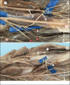
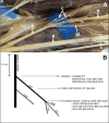
Results: The AIN originated from the median nerve in an average of 5.2 cm distal to the intercondylar line. In 29 limbs, it originated from the posterior fascicles of the median nerve, while in 21 specimens, from the posterolateral fascicles. In 2 specimens, two branches for the AIN were present. The PIN was studied in 30 limbs, we identified its origin in the radial nerve in all limbs. In 23 limbs, the branches to the supinator muscle originated from the PIN proximally to the Fröhse arcade, in 7 members distally.
Conclusion: The PQB was reliably present in all dissected forearms and presented variations only in its diameter. The PQB could be transferred to PIN without tension in all specimens even with full range of motion of the forearm. Level of Evidence IV; Case Series.
Objetivos: avaliar as características e variações anatômicas do nervo interósseo anterior (NIA) e definir a viabilidade da transferência do ramo pronador quadrado (RPQ) para o nervo interósseo posterior (NIP) sem tensão.
Materiais e métodos: Cinquenta membros superiores de 25 cadáveres adultos do sexo masculino foram dissecados, 20 foram preparados e cinco eram cadáveres frescos.
Resultados: O NIA originou-se do nervo mediano em média 5,2 cm distal à linha intercondilar. Em 29 membros originou-se dos fascículos posteriores do nervo mediano, enquanto em 21 espécimes, dos fascículos póstero-laterais. Em 2 espécimes, estavam presentes dois ramos para o NIA. O NIP foi estudado em 30 membros, identificamos sua origem no nervo radial em todos os membros. Em 23 membros, os ramos para o músculo supinador originaram-se do NIP proximalmente à arcada de Fröhse, em 7 membros distalmente.
Conclusão: O RPQ esteve presente de forma confiável em todos os antebraços dissecados e apresentou variações apenas em seu diâmetro. O RPQ pode ser transferido para NIP sem tensão em todos os espécimes, mesmo com amplitude total de movimento do antebraço. Nível de Evidência IV; Série de Casos.
Keywords: Cadaver; Dissection; Forearm; Muscles; Reconstructive Surgical Procedures.
Conflict of interest statement
All authors declare no potential conflict of interest related to this article.
Figures


References
LinkOut - more resources
Full Text Sources
