Review
. 2025 Jun 19;5(4):297-317.
doi: 10.1515/mr-2024-0095. eCollection 2025 Aug.

Spatio-temporal processes in autophagosome-lysosome fusion

Affiliations
Review

Spatio-temporal processes in autophagosome-lysosome fusion

Shizuo Liu et al. Med Rev (2021). .

Abstract

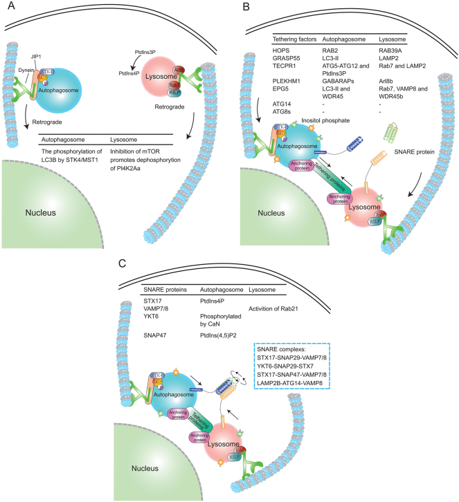
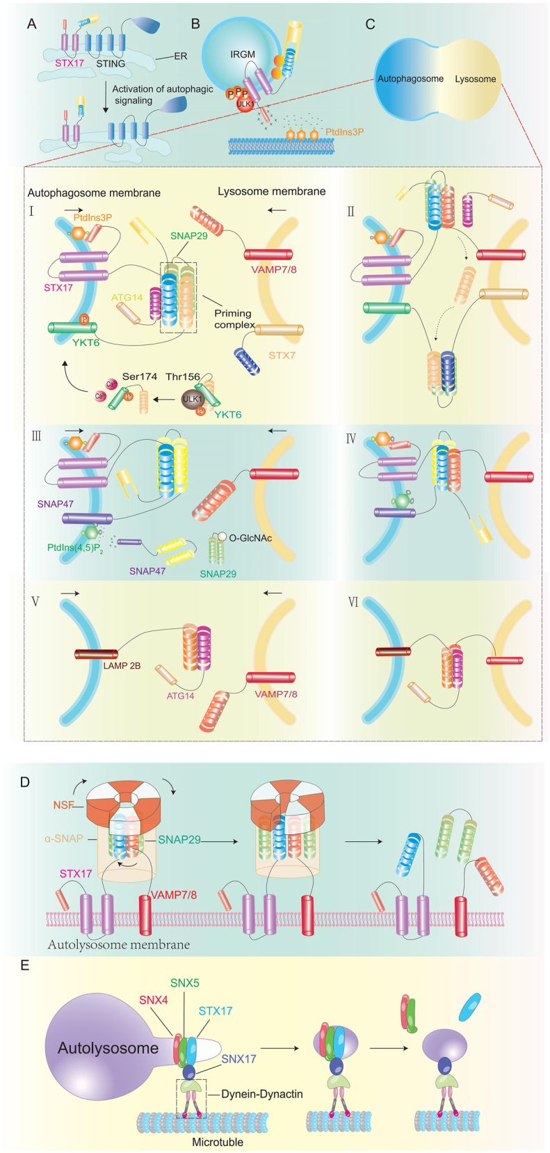
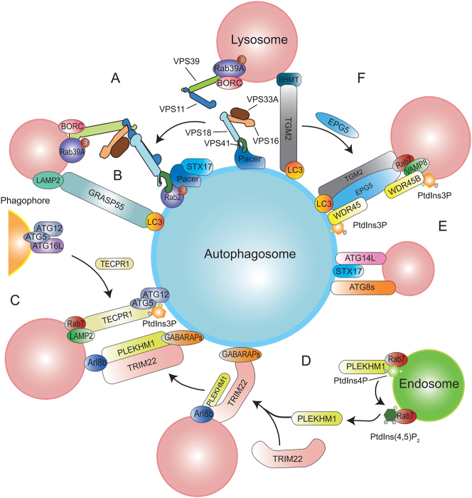
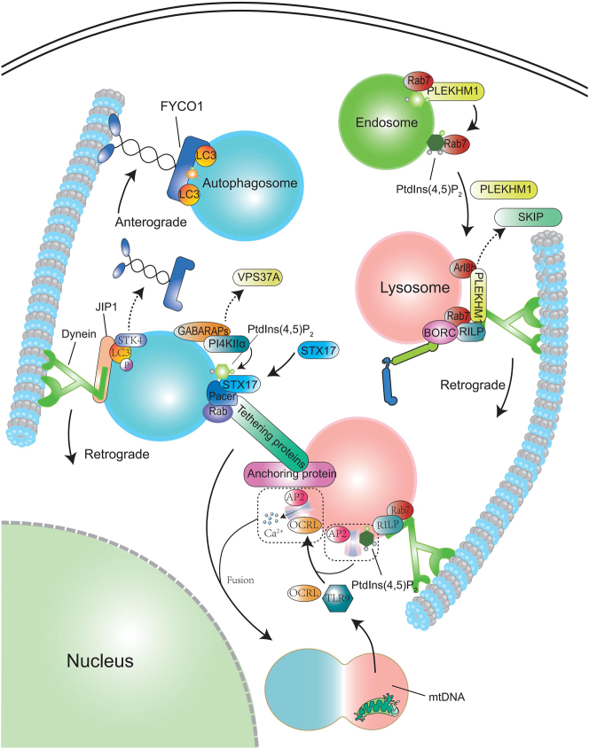
Macroautophagy/autophagy is a lysosome-dependent degradation process involved in cellular energy metabolism, recycling and quality control. Autophagy is a highly dynamic and precisely regulated process, which contains four major steps: autophagic membrane initiation and cargo recognition, autophagosome formation, autophagosome-lysosome fusion and lysosomal degradation. During the terminal phase of autophagy, the merging of the autophagosome and lysosome membranes is critical for the effective breakdown of sequestered cargoes. However, the participated molecules and the interplay among them have not been fully uncovered. The spatiotemporal property of these molecules is crucial for maintaining the orderly fusion of autophagosomes and lysosomes, otherwise it may lead to fusion disorders. In this article, we tend to summarize the molecules mediating autophagosome-lysosome fusion into two categories: effector molecules and regulatory molecules. The effector molecules are soluble N-ethylmaleimide-sensitive factor attachment protein receptor and tethering proteins, and the latter category contains phosphatidylinositol, Rab GTPases and ATG8-family proteins. The spatio-temporal properties of these autophagosome-lysosome fusion mediating molecules will be featured in this review.

Keywords: autophagosome-lysosome fusion; autophagy; sensitive factor attachment protein receptor; syntaxin 17.


Conflict of interest statement

Conflict of interest: The authors state no conflict of interest.

Figures

Figure 1:
Figure 1:
The process of autophagosome-lysosome fusion. a) Autophagosomes surrounding organelles and lysosomes localized at the cell periphery move through the cytoskeleton to the vicinity of the perinuclear area. b) Autophagosomes and lysosomes clustered at the cell periphery approach each other in the presence of tethering factors. c) When the two are sufficiently close together, autophagosomes and lysosomal membrane-localized SNARE proteins interact with each other to form a helical complex, which further pulls them in distance until fusion. We have tabulated the key signals for lysosomal and autophagosomal translocation to the perinucleus, and for the localization of tethering factors and SNARE proteins to the corresponding membranes.
Figure 2:
Figure 2:
Effector molecule-mediated autophagosome-lysosome fusion. A, B: autophagic translocation of STX17. STX17 is initially immobilized by STING distributed on the ER and released upon energetic stress. STX17 remains in an autoinhibitory conformation until it binds to SNAP29, the Habc structural domain being able to sequester SNARE. B: Free STX17 forms an ARP and is transported to the vicinity of the autophagosome membrane. The positively charged lysine and arginine at the C-terminus of STX17 are attracted to the negatively charged PtdIns4P localized on the autophagosome membrane. C: Four sets of complexes mediating autophagosome fusion. C-Ⅰ: STX17 distributes to the autophagosome membrane and first participates in the priming complex with SNAP29-YKT6. The helical structural domain of ATG14 also binds to the SNARE structural domain of STX17. C-Ⅱ: Upon proximity of the autophagosome to the lysosome, YKT6 is displaced by VAMP7/8, resulting in the assembly of STX17- SNAP29-VAMP7/8. Displaced YKT6 participates in the formation of YKT6-SNAP29-STX7. C-Ⅲ, Ⅳ: Under basal conditions, SNAP29 is O-GlcNAc-modified and unable to bind to STX17. Positively charged SNAP47 is attracted to the negatively charged PtdIns(4,5)P2 on the phagophore membrane and binds to the SNARE structural domain of STX17. STX17-SNAP47-VAMP7/8 is formed when the membranes are in close proximity to each other. C-Ⅴ, Ⅵ: LAMP2B-ATG14-VAMP7/8 mediates autophagosome fusion in cardiomyocytes. D: Depolymerization of the SNARE complex. STX17-SNAP29-VAMP7/8 is initially immobilized by α-SNAP and then binds to NSF. NSF’s rotational motion causes the SNARE complex to unhelix. E: STX17 condensed vesicles bud from autolysosomes with SNX4 and SNX5. SNX17 acts as a linker to connect SNX4-SNX5 to dynamin-dynamics. SNX4 and SNX5 are then disassembled from the vesicle. STX, syntaxin; SNARE, sensitive factor attachment protein receptor; SNX, sorting nexin.
Figure 3:
Figure 3:
Tethering proteins in autophagosome-lysosome fusion. A. Quaternary HOPS (VPS33A-VPS16-VPS18-VPS41) and binary HOPS (VPS39-VPS11) and are anchored to Rab2 on the autophagosome and Rab39A on the lysosome, respectively. Then, assembled into complete HOPS complexes. B. GRASP55 is anchored to autophagosomes and lysosomes via LC3 and LAMP2, respectively. C. TECPR1 competes with ATG16 for ATG5-ATG12. On autophagosomes, ATG5-ATG12 is involved in TECPR1-anchoring with PI3P. Rab7 is involved in lysosomal end anchoring of LAMP2. D. PLEKHM1 initially binds to Rab7 on the endosome. Upon conversion of PtdIns4P to PtdIns(4,5)P2 on endosomes, PLEKHM1 is released by Rab7. Then, PLEKHM1 may bind to Arl8b on lysosomes. TRIM22 promotes the binding of PLEKHM1 to GABARAPs on autophagosomes. E. ATG14 and ATG8s participate in tethering. F. EPG5 first binds to TGM2. LC3, WDR45 and PI3P are involved in the anchoring of EPG5 at the autophagosome end. Rab7, VAMP8, WDR45B and PI3P are involved in the anchoring at the lysosome end. HOPS, homotypic fusion and protein sorting; TECPR1, tectonin β-propeller repeat containing 1; TGM2, Transglutaminase 2; GABARAP, gamma-aminobutyric-acid-type-A-receptor-associated protein.
Figure 4:
Figure 4:
The in-cell spatial regulation of autophagosome-lysosome fusion. Autophagosomes are initially transported by FYCO1 to move away from the perinuclear area. After fusion conditions are exhibited, STK4 phosphorylates LC3 to dissociate from FYCO1. Movement towards the perinucleus is then mediated by JIP1. Endosomal conversion of PtdIns4P to PtdIns(4,5)P2 promotes PLEKHM1 translocation to the lysosome, allowing lysosomal movement towards the perinucleus. GABARAP may dissociate from VPS37A after autophagosome closure, and then activate PI4KIIα to produce PtdIns4P. PtdIns4P attracts STX17, which recruits thrombospondins and forms the SNARE complex. Upon fusion of autophagosomes with lysosomes, degraded mitochondria expose mitochondrial DNA. MtDNA activates TLR9, which promotes OCRL recruitment to lysosomes. OCRL activates TRPML1 to release Ca2+ and promote autophagosome-lysosome fusion. STX, syntaxin; GABARAP, gamma-aminobutyric-acid-type-A-receptor-associated protein.

Similar articles

References

    1. Yamamoto H, Zhang S, Mizushima N. Autophagy genes in biology and disease. Nat Rev Genet. 2023;24:382–400. doi: 10.1038/s41576-022-00562-w. - DOI - PMC - PubMed
    1. Jensen LE, Rao S, Schuschnig M, Cada AK, Martens S, Hummer G, et al. Membrane curvature sensing and stabilization by the autophagic LC3 lipidation machinery. Sci Adv. 2022;8:eadd1436. doi: 10.1126/sciadv.add1436. - DOI - PMC - PubMed
    1. Kim HJ, Li Q, Tjon-Kon-Sang S, So I, Kiselyov K, Soyombo AA, et al. A novel mode of TRPML3 regulation by extracytosolic pH absent in the varitint-waddler phenotype. Embo J. 2008;27:1197–205. doi: 10.1038/emboj.2008.56. - DOI - PMC - PubMed
    1. Ke PY. Molecular mechanism of autophagosome-lysosome fusion in mammalian cells. Cells. 2024;13 doi: 10.3390/cells13060500. - DOI - PMC - PubMed
    1. Zhu Y, Liu F, Jian F, Rong Y. Recent progresses in the late stages of autophagy. Cell Insight. 2024;3:100152. doi: 10.1016/j.cellin.2024.100152. - DOI - PMC - PubMed

LinkOut - more resources