

Assessment of Small Vessel Density Changes in Cerebral Autosomal Dominant Arteriopathy With Subcortical Infarcts and Leukoencephalophy (CADASIL) by High-Resolution Black-Blood MRI
- PMID: 40838861
- PMCID: PMC12462767
- DOI: 10.1002/jmri.70096
Assessment of Small Vessel Density Changes in Cerebral Autosomal Dominant Arteriopathy With Subcortical Infarcts and Leukoencephalophy (CADASIL) by High-Resolution Black-Blood MRI
Abstract
Background: Direct assessments of cerebral small vessels in Cerebral Autosomal Dominant Arteriopathy with Subcortical Infarcts and Leukoencephalopathy (CADASIL) remain a challenge.
Purpose: To investigate changes of cerebral small vessels in CADASIL using iso-0.5 mm black-blood MRI.
Study type: Case control study.
Population: Thirty-six genetically confirmed CADASIL patients (23 female, 43 ± 12.48 years) and 35 matched healthy controls (27 female, 40 ± 11.57 years).
Field strength/sequence: 3T using a T1-weighted turbo spin-echo with variable flip angles sequence.
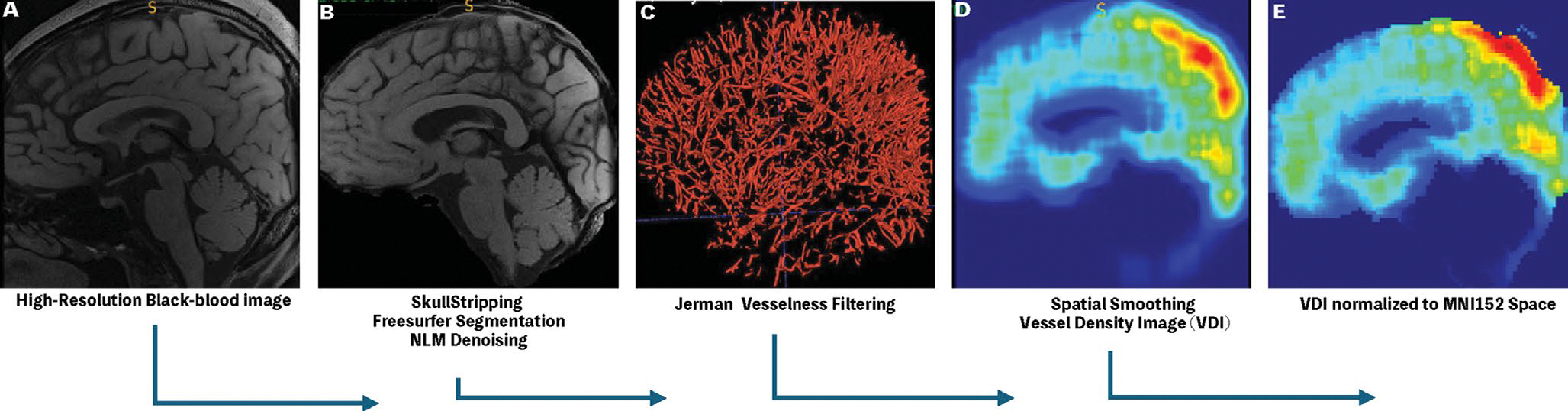
Assessment: Vessel density images (VDIs) were derived from black-blood MRI by using a semi-automatic pipeline with a Jerman filter. The differences in VDI were assessed between CADASIL and control groups. The relationships between changes in VDI and cognitive performance and disease burden were studied in the CADASIL group.
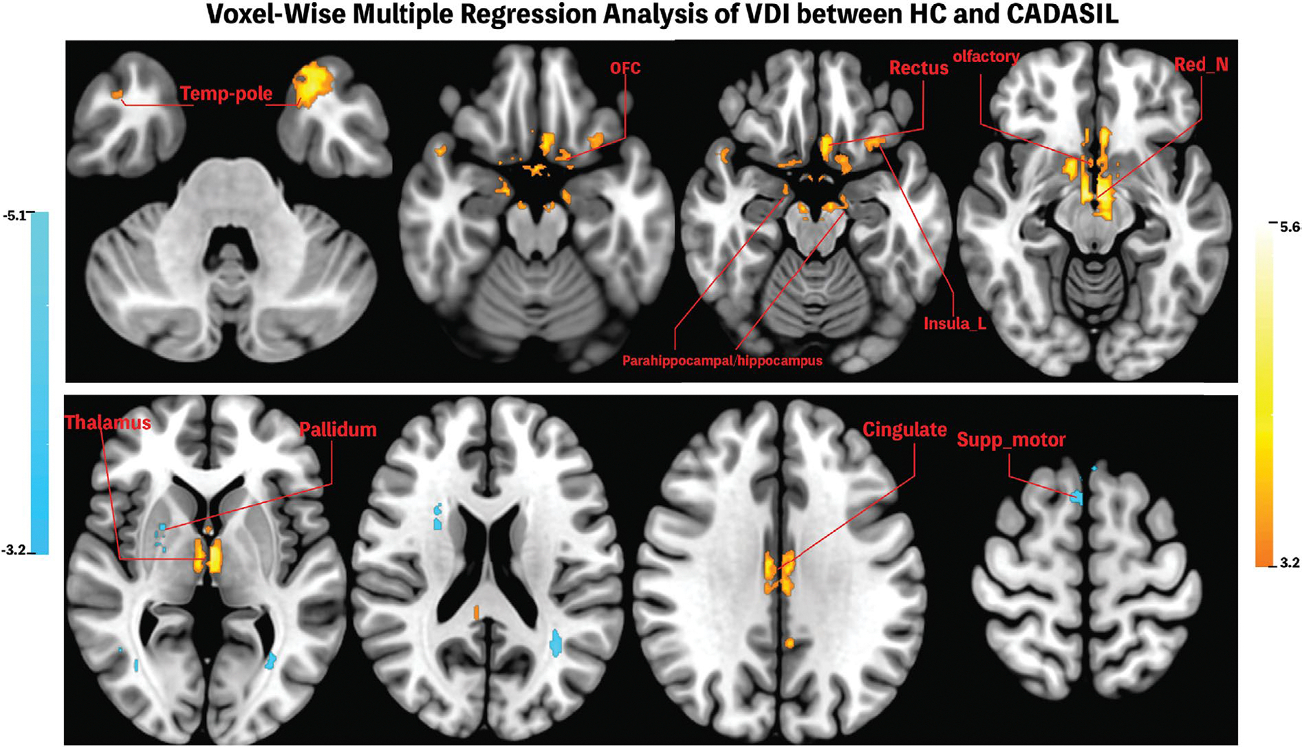
Statistical tests: Two-tailed independent samples t-tests were employed to assess the difference in VDI between CADASIL and control groups. Generalized linear mixed-effect models were used to assess the associations of VDI with cognitive performance and disease burden. Voxel-wise analyses were performed to further explore the associations of regional VDI with cognitive performance and disease burden after FDR correction.
Results: Reduced mean VDI was found in gray matter of CADASIL patients (1.31 ± 0.06) compared to controls (1.35 ± 0.03), which was significantly associated with lower MoCA scores (β = 52.89, SE = 12.99, 95% CI [26.38, 79.40]), and higher cerebral small vessel disease (cSVD) burden scores (β = -14.34, SE = 3.22, 95% CI [-20.91, -7.76]) in CADASIL patients. Voxel-wise analyses revealed reduced regional VDI in regions of the temporal pole, insula, cingulate cortex, and orbitofrontal cortex in CADASIL patients.
Data conclusion: The VDI technique based on high-resolution black-blood MRI demonstrated changes in regional VDI in CADASIL patients and offers a noninvasive imaging tool to advance the understanding of the mechanisms underlying cSVD.
Evidence level: 3.
Technical efficiency: Stage 2.
Keywords: Black‐blood MRI; CADASIL; cerebral small vessel disease; vessel density imaging.
© 2025 The Author(s). Journal of Magnetic Resonance Imaging published by Wiley Periodicals LLC on behalf of International Society for Magnetic Resonance in Medicine.
Figures


References
Grants and funding
LinkOut - more resources
Full Text Sources
