. 2025 Sep 1;5(9):1552-1565.
doi: 10.1158/2767-9764.CRC-25-0209.

Mesenchymal Stem Cells Expressing CES1 and Soluble TRAIL Activate CPT-11 and Induce Apoptosis in Lung Cancer Brain Metastatic Lesions

Affiliations

Mesenchymal Stem Cells Expressing CES1 and Soluble TRAIL Activate CPT-11 and Induce Apoptosis in Lung Cancer Brain Metastatic Lesions

Dong Oh Kim et al. Cancer Res Commun. .

Abstract

We aimed to develop a novel therapeutic strategy for lung cancer brain metastases by leveraging the tumor-tropic properties of genetically engineered Wharton's Jelly-derived mesenchymal stem cells (WJ-MSC) as vehicles for dual-agent gene therapy across the blood-brain barrier. WJ-MSCs were transiently engineered using lipid nanoparticle technology to coexpress soluble TRAIL (sTRAIL) and the prodrug-activating enzyme carboxylesterase 1 (CES1). In vitro analyses assessed transfection efficiency, therapeutic protein expression, apoptosis induction, and maintenance of stemness. Tumor-homing capacity was evaluated via EGFP labeling and intracerebral tracking. Therapeutic efficacy was tested in subcutaneous and intracerebral lung cancer xenograft models using bioluminescent imaging, histopathology, and IHC. In vivo treatment included intraperitoneal CPT-11 administration to assess synergy between CES1-mediated prodrug activation and sTRAIL-induced apoptosis. Modified WJ-MSCs exhibited preserved stem cell characteristics and strong tropism toward brain tumor sites. They secreted high levels of functional sTRAIL and CES1, enabling local activation of CPT-11 into SN-38 and inducing apoptosis through death receptor signaling (DR4/DR5). Combination therapy with WJ-MSCs-CES1.sTRAIL and CPT-11 significantly suppressed tumor growth in lung cancer brain metastasis models compared with control groups. The approach demonstrated selective cytotoxicity, minimal off-target effects, and favorable safety profiles. This study establishes a nonviral, transient gene delivery platform using autologous WJ-MSCs for dual-action gene therapy in lung cancer brain metastases. The combined use of CES1 and sTRAIL and enables precise tumor targeting and drug activation, offering a promising avenue for personalized, stem cell-based treatment strategies to improve outcomes in patients with brain metastatic lung cancer.

Significance: This study presents a nonviral, stem cell-based therapy for brain metastatic non-small cell lung cancer using WJ-MSCs expressing sTRAIL and CES1. These engineered cells home to tumors, activate CPT-11, and induce apoptosis. The dual-action strategy significantly reduced brain tumor burden with minimal toxicity, demonstrating strong therapeutic potential.


Conflict of interest statement

No disclosures were reported.

Figures

Figure 1.
Figure 1.
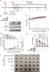
In vivo of therapeutic efficacy of mRNA-CES1 and sTRAIL for H460-Luc. A, Schematic illustrates an in vivo experiment. B, Bioluminescence imaging was performed using IVIS Optical Imaging equipment during the experiment. C, Representative subcutaneous tumors in the control and gene-transfected treatment groups. D, Graphs depicting the changes in tumor volume for each group. E, Graphs showing the changes in tumor weight for each group. All results are expressed as the mean ± SD; N = 4 for all groups. P values of statistical significance are represented as *, P < 0.05; **, P < 0.001; and ***, P < 0.000.
Figure 2.
Figure 2.
In vivo of therapeutic efficacy of LNP-mRNA-MSCs for H460-Luc. A, Schematic representation of an in vivo experiment. B, Confirming CES1 and FLT-3L expression in Western blot results of WJ-MSCs transfected with LNP-mRNA. C, A graph shows changes in the body weight of nude mice during WJ-MSCs-mRNA treatment. D, Average tumor volume. E, Average tumor weight. NS, not significant. F, Representative subcutaneous tumors in the control and treatment groups. All results are expressed as the mean ± SD; N = 6 for all groups. P values of statistical significance are represented as *, P < 0.05; **, P < 0.001; and ***, P < 0.0001.
Figure 3.
Figure 3.
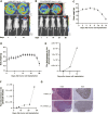
Modeling lung cancer brain metastasis using NSCLC. A and B, Tumor growth was monitored with luciferase expression via IVIS Spectrum imaging at different time points, and mice were imaged 10 minutes after luciferin intraperitoneal injection. C and D, Changes in mouse body weight after NSCLC injection. E and F, Plot of bioluminescence signal changes showing in vivo tumor growth. G, Histologic analysis of tumor tissues included hematoxylin and eosin (H&E) and Ki67 staining. Scale bar, 100 μm. All results are expressed as the mean ± SD.
Figure 4.
Figure 4.
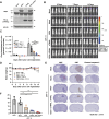
Therapeutic effects of treatment genes on lung cancer brain metastasis in vivo. A, Confirming CES1 and FLT-3L expression in Western blot results of H460-Luc cells transfected with LNP-mRNA. B, Bioluminescence imaging was conducted using IVIS Optical Imaging equipment during the experiment. The bioluminescent image of the first mouse in the mock day 7 condition is also presented for the day 7 condition in Fig. 3A, as the images represent the same experimental conditions. C, Bioluminescent signals were quantified using the IVIS imaging system, and results were obtained at 7 and 14 days after injecting H460-Luc cells for each group. D, Mouse body weight changes after injecting H460-Luc cells with transfection were assessed in each group of nude mice. E, Histologic analysis of tumor tissues in the H460-Luc model, stained with hematoxylin and eosin (H&E), Ki67, and cleaved caspase-3. F, A graph illustrating the area of cancer lesions in the mouse brain. All results are expressed as the mean ± SD; N = 5 for all groups. P values of statistical significance are represented as *, P < 0.05; **, P < 0.001; and ***, P < 0.0001.
Figure 5.
Figure 5.
Therapeutic tumor-homing ability of MSCs. A, Schematic representation of an in vivo experiment. B, Confirming EGFP expression in Western blot results of WJ-MSCs transfected with LNP-mRNA. C, The CLSM results depict brain cross-sections from 0 to 48 hours after ICV injection of WJ-MSCs-mRNA-EGFP into mouse brains. (Green represents Alexa Fluor 488 fluorescence, and blue represents DAPI staining). CLSM, confocal laser scanning microscope; DAPI, 4′,6-diamidino-2-phenylindole, dihydrochloride.
Figure 6.
Figure 6.
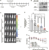
Therapeutic efficacy of CES1 and sTRAIL mRNAs delivered via WJ-MSCs in a lung cancer brain metastasis model. A, Schematic overview of the in vivo experimental design. B, Western blot analysis of CES1 and FLT-3L expression in WJ-MSCs transfected with LNP-encapsulated mRNAs. C, Bioluminescence imaging of tumor-bearing mice performed using the IVIS imaging system. D, Quantification of bioluminescent signals at 14 days after H460-Luc cell injection. NS, not significant. E, Body weight monitoring of nude mice throughout the treatment period. Data are presented as the mean ± SD; n = 5 per group. P values of statistical significance are represented as *, P < 0.05; **, P < 0.001.
Figure 7.
Figure 7.
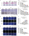
Histologic and molecular analyses of therapeutic outcomes in the brain metastasis model. A, Histologic examination of brain tumor tissues stained with hematoxylin and eosin (H&E) and Ki67. B, Quantification of tumor lesion area in the brain. C, Representative images of Ki67 immunostaining in brain tumor lesions. D, Quantification of Ki67-positive cells per group. E–H, Immunofluorescence staining for cleaved caspase-3 and CD31 in brain tissues, visualized using the ScanScope AT system (Leica Biosystems). Immunofluorescence staining for cleaved caspase-3 and CD31 in brain tissues, visualized using the ScanScope AT system (Leica Biosystems). E and F, Representative images and quantification of cleaved caspase-3–positive cells. G and H, Representative images and quantification of CD31-positive cells. Data are presented as the mean ± SD; n = 5 per group. P values of statistical significance are represented as *, P < 0.05; **, P < 0.001; and ***, P < 0.0001. DAPI, 4′,6-diamidino-2-phenylindole, dihydrochloride.

References

    1. Lee JG, Kim HC, Choi C-M. Recent trends of lung cancer in Korea. Tuberc Respir Dis (Seoul) 2021;84:89–95. - PMC - PubMed
    1. Siegel RL, Giaquinto AN, Jemal A. Cancer statistics, 2024. CA Cancer J Clin 2024;74:12–49. - PubMed
    1. Siegel RL, Miller KD, Jemal A. Cancer statistics, 2020. CA Cancer J Clin 2020;70:7–30. - PubMed
    1. Yang P. Epidemiology of lung cancer prognosis: quantity and quality of life. Methods Mol Biol 2009;471:469–86. - PMC - PubMed
    1. Nayak L, Lee EQ, Wen PY. Epidemiology of brain metastases. Curr Oncol Rep 2012;14:48–54. - PubMed

MeSH terms