. 2025 Aug 21;10(19):e184487.
doi: 10.1172/jci.insight.184487. eCollection 2025 Oct 8.

GABAergic interneurons contribute to the fatal seizure phenotype of CLN2 disease mice

Affiliations

GABAergic interneurons contribute to the fatal seizure phenotype of CLN2 disease mice

Keigo Takahashi et al. JCI Insight. .

Abstract

The cellular etiology of seizures in CLN2 disease, a childhood-onset neurodegenerative lysosomal storage disorder caused by a deficiency of tripeptidyl peptidase 1 (TPP1), remains elusive. Given that Cln2R207X/R207X mice display fatal spontaneous seizures and an early loss of several cortical GABAergic interneuron populations, we hypothesized that these 2 events might be causally related. To study the cell-autonomous effects of interneuron-specific TPP1 deficiency, we first generated transgenic mice expressing loxP-flanked lysosomal membrane-tethered TPP1 (TPP1LAMP1 mice) on the Cln2R207X/R207X genetic background, and then crossed TPP1LAMP1 mice with Vgat-Cre mice. These Vgat-Cre; TPP1LAMP1 mice accumulated storage material in cortical and striatal interneurons. Vgat-Cre; TPP1LAMP1 mice also died more readily after pentylenetetrazole-induced seizures, indicating that interneuron-specific TPP1 deficiency renders these mice more susceptible to seizure-induced mortality. We also selectively activated interneurons using designer receptors exclusively activated by designer drugs (DREADDs) in Vgat-Cre; Cln2R207X/R207X mice. Electroencephalogram monitoring revealed that DREADD-mediated activation of interneurons markedly accelerated the onset of spontaneous seizures and seizure-associated death in Vgat-Cre; Cln2R207X/R207X mice, suggesting that modulating interneuron activity can exacerbate epileptiform abnormalities. Taken together, these results provide mechanistic insights into the underlying etiology of seizures and premature death that characterize CLN2 disease.

Keywords: Genetic diseases; Genetics; Lysosomes; Neuroscience; Seizures.


Conflict of interest statement

Conflict of interest: JDC has received research support from BioMarin Pharmaceutical Inc., Abeona Therapeutics Inc., REGENXBIO Inc., and Neurogene, and is a consultant for JCR Pharmaceutical.

Figures

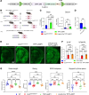
Figure 1
Figure 1. Generation of TPP1LAMP1 mice.
(A) The TPP1LAMP1 construct was created by linking human TPP1 cDNA with the transmembrane domain of LAMP1 via a 6-glycine linker under the PGK promoter. The entire construct was flanked by loxP loci. (B) Schematic describing the breeding strategy to generate TPP1LAMP1 mice. (C) TPP1 activity assays in the brain (left) at 15 weeks show supraphysiological TPP1 activity in TPP1LAMP1 mice vs. WT mice, and indistinguishable TPP1 activity in β-actin-Cre; TPP1LAMP1 mice from Cln2R207X/R207X mice (n = 4 per group). TPP1 activity assays (right) show supraphysiological TPP1 activity in the serum of TPP1LAMP1 mice compared with WT (n = 5 in WT and Cln2R207X/R207X mice and n = 4 in TPP1LAMP1 mice). (D) Binding to CI-M6PR is significantly reduced in TPP1LAMP1 compared with endogenous WT TPP1 in the brain (n = 4 per group). (E) Immunostaining for SCMAS (green) shows widespread SCMAS accumulation in Cln2R207X/R207X mice and β-actin-Cre; TPP1LAMP1 mice, but not in WT and TPP1LAMP1 mice at 15 weeks. Scale bar: 1 mm. (F) Quantitative analysis of SCMAS immunoreactivity in the S1BF and VPM/VPL at 15 weeks confirms that SCMAS accumulation seen in Cln2R207X/R207X mice (red bars) is completely rescued in TPP1LAMP1 mice (green bars) and is fully recapitulated in β-actin-Cre; TPP1LAMP1 mice (purple bars) across multiple brain regions (n = 6 per group). (G) CatWalk XT gait analysis shows significantly shorter stride length and swing duration, wider distance between hind paws (base of support or BOS), and higher proportion of steps supported by 3 feet in 15-week-old Cln2R207X/R207X mice (red) compared with age-matched WT mice (blue). These gait abnormalities were rescued in TPP1LAMP1 mice (green) and recapitulated in β-actin-Cre; TPP1LAMP1 mice (purple) at the same age (n = 10 in WT, Cln2R207X/R207X, and TPP1LAMP1 mice and n = 9 in β-actin-Cre; TPP1LAMP1). Dots represent values from individual animals. Values are shown as mean ± SEM. One-way ANOVA with Bonferroni’s correction (C, F, and G) and unpaired, 2-tailed t test (D). *P < 0.05; **P < 0.01; ***P < 0.001; ****P < 0.0001.
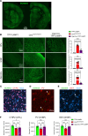
Figure 2
Figure 2. Interneuron-specific TPP1 deficiency leads to storage material accumulation and interneuron loss.
(A) Immunostaining for SCMAS (green) in the brain of Vgat-Cre; TPP1LAMP1 at 15 weeks of age shows scattered distribution of SCMAS-positive cells across the brain. (B) Comparison of immunostained brain sections for SCMAS (green) and quantitative analysis of their immunoreactivity via thresholding image analysis between TPP1LAMP1, Cln2R207X/R207X, and Vgat-Cre; TPP1LAMP1 mice across multiple brain regions reveals partial storage material accumulation in Vgat-Cre; TPP1LAMP1 compared with Cln2R207X/R207X mice, with statistically significant differences observed within the CPu at 15 weeks of age. (C) Coimmunostaining for SCMAS (green), CTIP2 (red), and Nissl (cyan) reveals storage material accumulation in a subset of CTIP2-positive MSNs within the CPu in Vgat-Cre; TPP1LAMP1 mice. (D) Coimmunostaining for SCMAS (green) and PV (red) reveals storage material accumulation in a subset of PV-positive interneurons within the S1BF in Vgat-Cre; TPP1LAMP1 mice. (E) Coimmunostaining for SCMAS (green), SST (red), and Nissl (cyan) reveals storage material accumulation in a subset of SST-positive interneurons within the S1BF cortex in Vgat-Cre; TPP1LAMP1 mice. (F) Unbiased stereological counts of immunostained neuron populations within the CPu reveals a significant loss of CTIP2-positive medium spiny neurons in Vgat-Cre; TPP1LAMP1 mice to a comparable extent to that in Cln2R207X/R207X mice at 15 weeks of age. The same unbiased stereological analysis within the S1BF cortex reveals no significant loss of PV- or SST-positive neurons in Vgat-Cre; TPP1LAMP1 mice compared to those in TPP1LAMP1 mice at 15 weeks of age. Dots represent values from individual animals. Values are shown as mean ± SEM (n = 6 mice per group). One-way ANOVA with Bonferroni’s correction. *P < 0.05; **P < 0.01; ***P < 0.001; ****P < 0.0001. Scale bars: 1 mm (A), 200 μm (B), and 50 μm (CE).
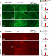
Figure 3
Figure 3. Interneuron-specific TPP1 deficiency does not trigger the neuroimmune response associated with CLN2 disease.
Immunostaining for GFAP (A, green) and CD68 (B, red) and quantitative analysis of their immunoreactivity via thresholding image analysis in the CPu, S1BF, and VPM/VPL at 15 weeks of age shows the marked increase in the intensity of GFAP and CD68 immunoreactivities in Cln2R207X/R207X mice (red bars), but no significant increase in Vgat-Cre; TPP1LAMP1 mice (purple bars) compared to age-matched TPP1LAMP1 mice (green bars). Dots represent values from individual animals. Values are shown as mean ± SEM (n = 6 mice per group). One-way ANOVA with Bonferroni’s correction. *P < 0.05; **P < 0.01; ***P < 0.001; ****P < 0.0001. Scale bars: 200 μm.
Figure 4
Figure 4. Interneuron-specific TPP1 deficiency increases susceptibility to sudden death secondary to pentylenetetrazole-induced seizures.
(A) CatWalk XT gait analysis reveals preserved gait performance of Vgat-Cre; TPP1LAMP1 mice (purple) compared with TPP1LAMP1 and Cln2R207X/R207X mice at 15 weeks of age (n = 10 per group). (B) Seizures were induced by intraperitoneal injection of pentylenetetrazole (PTZ; 75 mg/kg) in TPP1LAMP1 (n = 12, green bars) and Vgat-Cre; TPP1LAMP1 (n = 9, purple bars) mice at 25 weeks of age. There was no significant difference in the latency to tonic-clonic seizures between 2 groups. Vgat-Cre; TPP1LAMP1 mice exhibit a significantly lower number of tonic-clonic seizures per mouse and a significantly shorter time to death compared with TPP1LAMP1 mice. The average duration of nonfatal tonic-clonic seizures in TPP1LAMP1 mice was significantly shorter than the average duration of fatal tonic-clonic seizures in TPP1LAMP1 mice and Vgat-Cre; TPP1LAMP1 mice. Dots represents values from individual animals. Values are shown as mean ± SEM. One-way ANOVA with Bonferroni’s correction (A) and unpaired, 2-tailed t test (B). *P < 0.05; **P < 0.01; ***P < 0.001; ****P < 0.0001.
Figure 5
Figure 5. Chemogenetic activation of interneurons exacerbates seizure phenotypes in Cln2R207X/R207X mice.
(A) Schematic of experimental design. The image was created with BioRender.com. (B) Fluorescence images depict widespread transduction of intracerebroventricularly delivered AAV9-hSyn-DIO-hM3Dq-mCherry (red) in Vgat-Cre; Cln2R207X/R207X mouse brain at 6 weeks of age, predominantly in the cortex and striatum. Lower-magnification images (top) and higher-magnification images focused on the CPu and S1BF (bottom). Scale bars: 1 mm (top) and 200 μm (bottom). (C) Time course of seizures (red dots) and deaths (black Xs) in hM3Dq-mCherry–expressing (top) and mCherry-expressing (bottom) Vgat-Cre; Cln2R207X/R207X mice. EEG recordings revealed a significantly earlier onset of spontaneous seizures (D) and premature death (E) in hM3Dq-mCherry–expressing Vgat-Cre; Cln2R207X/R207X mice upon chronic deschloroclozapine (DCZ) administration compared with mCherry-expressing control mice (n = 8 per group). Log-rank (Mantel-Cox) test. (F) Representative EEG traces show an increased frequency of abnormal spikes (top) and burst-suppression activity (middle) in hM3Dq-mCherry–expressing Vgat-Cre; Cln2R207X/R207X mice during the first week of DCZ administration compared with mCherry-expressing Vgat-Cre; Cln2R207X/R207X mice (bottom).
Figure 6
Figure 6. Chemogenetic activation of interneurons alters microglial activation in Cln2R207X/R207X mice.
(A) Schematic of experimental design. The image was created with BioRender.com. (B) Immunostaining for GFAP (green) and CD68 (red) and quantitative analysis of their immunoreactivity via thresholding image analysis reveals a significantly reduced CD68 immunoreactivity in hM3Dq-mCherry–expressing Vgat-Cre; Cln2R207X/R207X mice (n = 5, red bars) upon DCZ administration across the CPu, S1BF, and VPM/VPL with no change in GFAP immunoreactivity compared to mCherry-expressing control mice (n = 6, gray bars). Scale bars: 200 μm. Dots represents values from individual animals. Values are shown as mean ± SEM. Unpaired, 2-tailed t test. ****P < 0.0001.
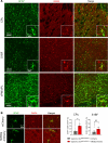
Figure 7
Figure 7. Chemogenetic activation of interneurons aggravates GABA immunoreactivity within astrocytes in Cln2R207X/R207X mice.
Coimmunostaining for GFAP (green) and GABA (red) shows overlap between the 2 channels across the CPu, S1BF, and VPM/VPL in 12-week-old Cln2R207X/R207X mice. Insets are higher-magnification views from each image. Scale bars: 100 μm (low magnification) and 20 μm (high magnification). (B) Immunostaining for GFAP (green) and GABA (red) and colocalization analysis on the confocal images reveals a significantly increased GABA immunoreactivity in GFAP-positive astrocytes within the CPu of hM3Dq-mCherry–expressing Vgat-Cre; Cln2R207X/R207X mice (n = 5, red bars) upon DCZ administration compared with mCherry-expressing control mice (n = 6, gray bars). There was a similar increase in the GABA immunoreactivity in GFAP-positive astrocytes within the S1BF, but it was not statistically significant. Scale bar: 20 μm. Dots represents values from individual animals. Values are shown as mean ± SEM. Unpaired, 2-tailed t test. *P < 0.05.

Update of

References

    1. Lee S, et al. A disinhibitory circuit mediates motor integration in the somatosensory cortex. Nat Neurosci. 2013;16(11):1662–1670. doi: 10.1038/nn.3544. - DOI - PMC - PubMed
    1. Pi HJ, et al. Cortical interneurons that specialize in disinhibitory control. Nature. 2013;503(7477):521–524. doi: 10.1038/nature12676. - DOI - PMC - PubMed
    1. Canto-Bustos M, et al. Disinhibitory circuitry gates associative synaptic plasticity in olfactory cortex. J Neurosci. 2022;42(14):2942–2950. doi: 10.1523/JNEUROSCI.1369-21.2021. - DOI - PMC - PubMed
    1. Staley K. Molecular mechanisms of epilepsy. Nat Neurosci. 2015;18(3):367–372. doi: 10.1038/nn.3947. - DOI - PMC - PubMed
    1. Armada-Moreira A, et al. Going the extra (synaptic) mile: excitotoxicity as the road toward neurodegenerative diseases. Front Cell Neurosci. 2020;14:90. doi: 10.3389/fncel.2020.00090. - DOI - PMC - PubMed

MeSH terms

Supplementary concepts