. 2025 Sep 22;35(18):4349-4364.e5.
doi: 10.1016/j.cub.2025.07.059. Epub 2025 Aug 20.

A circuit from the basolateral amygdala to hippocampal CA3 regulates social behavior

Affiliations

A circuit from the basolateral amygdala to hippocampal CA3 regulates social behavior

Minghua Li et al. Curr Biol. .

Abstract

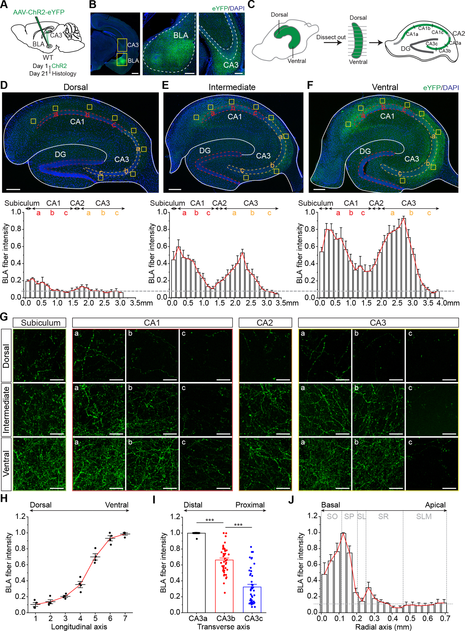
The CA3 region of the hippocampus is essential for associative memory. CA3 pyramidal neurons receive three canonical excitatory inputs-recurrent collaterals from other CA3 pyramidal neurons, mossy fiber input from the dentate gyrus (DG), and perforant path input from the entorhinal cortex-that terminate at specific dendritic compartments and have distinct functions. Yet, the additional extrahippocampal inputs to CA3 are less clear. Here, we report a monosynaptic glutamatergic input from the basolateral amygdala (BLA) that preferentially innervates ventral CA3. The CA3-projecting BLA neurons are topographically clustered in a small area near the medial border of BLA and preferentially innervate basal dendrites of distal CA3 (near CA1). Moreover, the BLA input preferentially excites regular-spiking CA3 pyramidal neurons expressing thorny excrescences, largely avoids burst-firing CA3 neurons lacking thorny excrescences (athorny cells), and only weakly excites CA2 pyramidal neurons. Furthermore, chemogenetic or optogenetic manipulations of the BLA-CA3 pathway bidirectionally alter social behavior. Taken together, our findings demonstrate that the BLA input constitutes a major glutamatergic input that can robustly excite CA3 pyramidal neurons in a cell-type- and subregion-specific manner and regulate social behavior.

Keywords: CA2; CA3; athorny; basolateral amygdala; hippocampus; memory; pyramidal neurons; social behavior; thorny excrescences.


Conflict of interest statement

Declaration of interests The authors declare no competing interests.

Figures

Figure 1.
Figure 1.. Topographical distribution of BLA fibers in the hippocampus.
(A) Experimental procedure and timeline. (B) Left, coronal section showing the injection site in BLA and BLA fibers in CA3. Enlarged images from the yellow boxes shown in the middle (BLA) and right (CA3). Scale bars, 500 μm (left), 200 μm (middle, right). (C) Experimental procedure showing that the hippocampus was dissected out and sectioned along the longitudinal axis. (D-F) Confocal images of the transverse sections from dorsal (D), intermediate (E), and ventral hippocampus (F). Bottom, quantification of BLA fibers along the transverse axis. Dashed line indicates the background noise. Scale bars, 200 μm. (G) Enlarged images of the yellow squares in each region in (D-F). Scale bars, 20 μm. (H-J) Group data of the normalized BLA fiber intensity along the longitudinal (H), transverse (I), and radial axes (J). Horizontal dashed line indicates the background noise. Vertical dashed lines indicate the borders of different stratums. ***p < 0.001. Error bars denote as s.e.m. See also Figures S1–S3.
Figure 2.
Figure 2.. Topographic heterogeneity of BLA neurons that project to vCA3 vs vCA1.
(A) Experimental procedure and timeline. (B-D) Confocal images of the injection sites in vCA3 (B) and vCA1 (C), and the AAVretro-labeled presynaptic neurons in BLA (D). Scale bars, 500 μm (B-C), 100 μm (D). (E) Distribution of vCA3-, vCA1-, and dual-projecting BLA neurons along the AP axis. (F) Line chart of the number of AAVretro-labeled cells in BLA along the AP axis (n = 3–4 mice for each group, two-way mixed ANOVA with post hoc Bonferroni test). (G) The estimated ratios of vCA3-, vCA1-, and dual-projecting BLA neurons relative to all BLA neurons [n = 4 for each group, one-way ANOVA, F(2, 9) = 200.43696, p = 3.44 × 10⁻⁸]. (H) Line chart of the number of AAVretro-labeled cells in BLA along the medial-lateral (ML) axis around Bregma −2.15 mm (n = 4 mice, 8 sections for each group, two-way mixed ANOVA with post hoc Bonferroni test). *p < 0.05; **p < 0.01; ***p < 0.001. Error bars denote as s.e.m. See also Figures S4.
Figure 3.
Figure 3.. CA3-projecting BLA neurons preferentially project to CA3 over other regions.
(A) Schematic indicating the site of AAVretro injection in CA3 and Cre-dependent ChR2 injection in BLA. Right, experimental timeline. (B) Sample images of AAVretro-labeled presynaptic neurons in BLA (red), some of which express ChR2-eYFP (green). Arrows show two mCherry+/eYFP+ cells. Scale bars, 50 μm (left), 20 μm (right). (C-E) Confocal images of the dissected transverse sections from dorsal (C), intermediate (D), and ventral hippocampus (E). Bottom, quantification of fibers of CA3-projecting BLA neurons along the transverse axis. Dashed line indicates the background noise. Scale bars, 200 μm. (F) Enlarged images of the yellow squares shown in (C-E). Scale bars, 20 μm. (G) Confocal images of mPFC, NAc and CeA from all BLA neurons (top) and CA3-projecting BLA neurons (bottom). Scale bars, 20 μm. (H) Quantification of BLA fiber intensity in extrahippocampal regions of all BLA neurons vs CA3-projecting BLA neurons (n = 3 for each group). *p < 0.05; **p < 0.01. Error bars denote as s.e.m See also Figures S5 and S6.
Figure 4.
Figure 4.. BLA input preferentially targets thorny pyramidal neurons in distal vCA3 and largely avoids athorny CA3 neurons.
(A) Experimental procedure and timeline. (B) Confocal images of biocytin-filled CA3a and CA3c neurons. Scale bars, 100 μm. (C) Sample traces (left) and group data (right) of oEPSCs recorded from CA3a and CA3c pyramidal neurons in voltage-clamp (n = 10 pairs, p = 0.0024, paired t-Test). Blue bar indicates light pulse. (D) Sample traces (left) and normalized group data (right) of oEPSCs in voltage-clamp in the absence and presence of DNQX (20 μM) and DL-APV (50 μM) (n = 6 for each group, p = 1.65 × 10⁻⁸, paired t-Test). (E) Group data of oEPSC onsets in response to 2-ms light stimulation. (F) Sample traces (left) and normalized group data (right) of oESPCs in voltage-clamp in baseline, after bath application of TTX (1 μM), and application of both TTX and 4-AP (100 μM) [n = 7 cells, p = 2.38 × 10⁻¹⁰, one-way ANOVA, F(2, 18) = 96.5561]. (G) Confocal images of biocytin-filled thorny and athorny cells. Scale bars, 100 μm, 10 μm (amplified images). (H) Distribution of the cell body positions of thorny and athorny cells along the radial axis in SP (n = 14 for each group). s, superficial; d, deep. (I) Sample traces of current-clamp recording in thorny and athorny CA3 cells in response to somatic current injections. (J) Firing frequency as a function of different steps of somatic current injections in thorny (n = 15) and athorny CA3 cells (n = 12, two-way mixed ANOVA with post hoc Bonferroni test). (K) Instantaneous firing frequency in response to 800-pA current injections in thorny (n = 9) and athorny cells (n = 7, two-way mixed ANOVA with post hoc Bonferroni test). (L) Sample traces (left) and group data (right) of oEPSCs recorded from thorny (n = 24) and athorny cells (n = 20) in voltage-clamp (p = 4.81 × 10⁻⁶, unpaired t-Test). (M) The probability of functional connections between BLA and thorny cells (100%, 24/24 cells) vs between BLA and athorny cells (10%, 2/20 cells). **p < 0.01; ***p < 0.001. Error bars denote as s.e.m.
Figure 5.
Figure 5.. BLA input weakly excites vCA2 pyramidal neurons.
(A) Left, confocal image of a ventral transverse section from a mouse injected with ChR2-eYFP into the BLA. Right, enlarged views of the boxes on the left. PCP4 is a marker for CA2. Scale bars, 200 μm (left), 50 μm (right). (B) Quantification of fluorescence intensity of PCP4 (red) and ChR2-eYFP (green) along the CA2-CA3 transverse axis. Shades depict s.e.m. (C) Confocal images of the biocytin-filled CA2 and CA3 pyramidal neurons. Scale bar, 100 μm. Expanded views show the absence and presence of thorny excrescences in CA2 and CA3 neurons, respectively. Scale bar, 20 μm. (D) Sample traces of current clamp recording of CA2 and CA3 neurons in response to different current injections. (E) Group data of input resistance of CA2 (n = 13) and CA3 cells (n = 8, p = 0.00907, unpaired t-Test). (F) Firing rate of CA2 (n = 9) and CA3 cells (n = 12) in response to step current injections (two-way mixed ANOVA with post hoc Bonferroni test). (G) Sample traces (left) and group data (right) of oEPSCs recorded from CA2 (n = 13) and CA3 cells (n = 8) in voltage-clamp (p = 4.68 × 10⁻⁴, unpaired t-Test). Blue bar indicates light pulse. (H) Sample traces (left) and group data (right) of optically-evoked PSPs (oPSPs) recorded in CA2 (n = 6) and CA3 cells (n = 9) in response to a train of 20-Hz light stimulation (n = 9, two-way mixed ANOVA with post hoc Bonferroni test). **p < 0.01; ***p < 0.001. Error bars denote as s.e.m.
Figure 6.
Figure 6.. Suppression of CA3-projecting BLA neurons alters social behavior.
(A) Experimental timeline. (B) Schematic (left) and representative images (right) of AAVretro-Cre-eGFP injection in CA3 and Cre-dependent hM4D or mCherry injection in BLA. Scale bar, 500 μm (left), 50 μm (right). (C) Experimental timeline of open field test. i.p., intraperitoneal injection. (D) Group data of distance traveled (left) and center time (right) in open field test. (E) Experimental timeline of social interaction test. (F) Group data of social interaction time (n = 18 for mCherry, n = 14 for hM4D, p = 0.0362, unpaired t-Test,). (G) Experimental timeline of three-chamber social memory test. (H) Left, interaction time with a mouse (M) or an object (O) (n = 16 for mCherry, n = 11 for hM4D, two-way mixed ANOVA with post hoc Bonferroni test). Right, discrimination index. (I) Left, social interaction time with a familiar mouse (FM) or a novel mouse (NM) (n = 16 for mCherry, n = 11 for hM4D, two-way mixed ANOVA with post hoc Bonferroni test). Right, discrimination index (n = 16 for mCherry, n = 11 for hM4D, p = 0.0921, unpaired t-Test). ns, not significant; *p < 0.05; **p < 0.01; ***p < 0.001. Error bars denote as s.e.m. See also Figures S7.
Figure 7.
Figure 7.. Stimulation of BLA terminals in CA3 alters social behavior.
(A) Experimental timeline. (B) Schematic (left) and representative images (right) of AAV-ChR2-eYFP or AAV-eYFP injection in BLA and fiber implanting in CA3. Scale bar, 500 μm (left), 200 μm (right). (C) Experimental timeline of open field test. (D) Group data of center time in open field test. (E) Experimental timeline of counterbalanced social interaction tests. (F) Time difference of social interaction between light-on and light-off (n = 8 for eYFP, n = 9 for ChR2, p = 0.00899, unpaired t-Test). (G) Experimental timeline of three-chamber social memory tests. (H) Left, interaction time with a mouse (M) or an object (O) with light delivered (n = 9 for eYFP, n = 8 for ChR2, two-way mixed ANOVA with post hoc Bonferroni test). Right, discrimination index (eYFP: n = 9; ChR2: n = 8, p = 0.06633, unpaired t-Test). (I) Left, interaction time with a mouse (M) or an object (O) without light stimulation (n = 9 for eYFP, n = 8 for ChR2, two-way mixed ANOVA with post hoc Bonferroni test). Right, discrimination index. (J) Left, social interaction time with a familiar mouse (FM) or a novel mouse (NM) with light delivered (n = 9 for eYFP, n = 8 for ChR2, two-way mixed ANOVA with post hoc Bonferroni test). Right, discrimination index (n = 9 for eYFP, n = 8 for ChR2, p = 0.03452, unpaired t-Test). ns, not significant; *p < 0.05; **p < 0.01; ***p < 0.001. Error bars denote as s.e.m. See also Figures S7.

References

    1. Rebola N, Carta M, and Mulle C (2017). Operation and plasticity of hippocampal CA3 circuits: implications for memory encoding. Nat Rev Neurosci 18, 208–220. 10.1038/nrn.2017.10. - DOI - PubMed
    1. Guzman SJ, Schlogl A, Frotscher M, and Jonas P (2016). Synaptic mechanisms of pattern completion in the hippocampal CA3 network. Science 353, 1117–1123. 10.1126/science.aaf1836. - DOI - PubMed
    1. Nakazawa K, Sun LD, Quirk MC, Rondi-Reig L, Wilson MA, and Tonegawa S (2003). Hippocampal CA3 NMDA receptors are crucial for memory acquisition of one-time experience. Neuron 38, 305–315. - PubMed
    1. Nakazawa K, Quirk MC, Chitwood RA, Watanabe M, Yeckel MF, Sun LD, Kato A, Carr CA, Johnston D, Wilson MA, and Tonegawa S (2002). Requirement for hippocampal CA3 NMDA receptors in associative memory recall. Science 297, 211–218. 10.1126/science.1071795. - DOI - PMC - PubMed
    1. Watson JF, Vargas-Barroso V, Morse-Mora RJ, Navas-Olive A, Tavakoli MR, Danzl JG, Tomschik M, Rossler K, and Jonas P (2024). Human hippocampal CA3 uses specific functional connectivity rules for efficient associative memory. Cell. 10.1016/j.cell.2024.11.022. - DOI

LinkOut - more resources