. 2025 Aug 21;42(5):49.
doi: 10.1007/s10585-025-10365-y.

Morphological heterogeneities in prostate cancer bone metastases are related to molecular subtypes and prognosis

Affiliations

Morphological heterogeneities in prostate cancer bone metastases are related to molecular subtypes and prognosis

Sofia Halin Bergström et al. Clin Exp Metastasis. .

Abstract

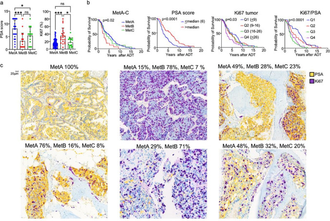
We previously identified three molecular subtypes of prostate cancer (PC) bone metastases, MetA-C, with MetB linked to poor prognosis after androgen deprivation therapy (ADT). This study analyzed epithelial and stromal markers using immunohistochemistry, focusing on their relationship to MetA-C subtypes, spatial heterogeneities, and clinical outcomes after ADT. High tumor proliferation and low PSA expression were associated with MetB and poor outcomes after ADT. Most metastases contained tumor epithelial subclones with different morphologies. In the metastasis stroma, blood vessels and fibroblast-like cells expressed smooth muscle actin (SMA), platelet-derived growth factor β, stroma-derived factor 1 (SDF1), periostin (POSTN), and decorin (DCN). Compared to each other, MetB metastases had higher SMA and ERG + endothelial cell densities, while MetA cases showed higher SDF1 and DCN levels. Accordingly, high POSTN and ERG + densities were associated with poor outcomes after ADT, whereas high DCN indicated favorable prognosis. Low levels of AR-positive stromal cells were linked to poor outcomes. Macrophage and T-lymphocyte densities showed no significant associations with metastases subtypes or outcome. Two stroma subtypes were identified: subtype 1 with higher bone content, lower vessel density, MetA-enrichment and better prognosis compared to subtype 2 that exhibited higher tumor proliferation and lower PSA expression. Most metastases contained regions of both stroma subtypes.

Keywords: Androgen deprivation therapy; Bone metastasis stroma; Metastases morphology; Metastatic stroma; Prostate cancer metastases; Prostate cancer molecular subtypes; Stromal markers; Tumor heterogeneity; Tumor microenvironment.


Conflict of interest statement

Declarations. Conflict of interest: A.B., E.T., and P.W. have a pending patent application related to the metastasis subtypes MetA-C and to Ki67 and PSA immunostaining as surrogate markers for the molecular metastasis subtypes (‘Methods for diagnosis and prognosis of prostate cancer’, EP2020/054681).

Figures

Fig. 1
Fig. 1
Tumor cell proliferation and PSA expression in relation to the molecular metastasis subtypes MetA-C and patient prognosis. a Graphs showing how tumor epithelial cell PSA and Ki67 staining scores differ between MetA, Met B and MetC dominating metastasis subtypes. * p < 0.05, *** p < 0.001, ns = not significantly different. b Kaplan-Meier curves showing survival after androgen-deprivation therapy (ADT) in relation to dominant molecular subtypes (MetA-C), tumor cell PSA, Ki67, and Ki67/PSA staining scores. c Sections from bone metastases, classified to contain different fractions of the MetA, MetB and MetC subtypes, double-stained for PSA and Ki67. Metastases with high MetA fractions (left) showed homogeneously strong PSA and low Ki67 staining, whereas metastases with high MetB fractions (middle) showed the opposite pattern. In metastases with a dominating MetA fraction but also containing a substantial MetB component (right), local areas with moderate to high PSA and low to moderate Ki67 (MetA-like pattern) and adjacent areas with low PSA and high Ki67 (MetB-like pattern) were seen, suggesting spatial heterogeneities in tumor cell phenotypes
Fig. 2
Fig. 2
Stroma cell heterogeneities in relation to the molecular metastasis subtypes MetA-C and patient prognosis. Immunohistochemical localization of factors expressed in a blood vessel walls and b fibroblast-like cells. c Double staining of ERG (localized in endothelial cell nuclei) and various stroma cell markers. All markers except DCN were localized adjacent to endothelial cells. d Double staining of DCN + SMA, and DCN + PDGFRB. SMA was expressed mainly in blood vessel walls adjacent to tumor epithelial cells whereas DCN was expressed in the broad stroma septa. PBGDRB was expressed adjacent to DCN in broad stroma septa and in blood vessels adjacent to tumor cells. e Multiplex staining showing that most stroma cell markers were expressed in the broad stroma septa whereas SMA, SDF-1 and focal PDGFRB were also detected in and around the tiny blood vessels penetrating tumor cell nests. f Kaplan-Meier analysis showing that survival after androgen-deprivation therapy (ADT) was related to the density of ERG + endothelial cells and to the stroma densities of POSTN and DCN. g Boxplots showing densities of stroma SMA, ERG, SDF-1, DCN, PDGFRB, and POSTN in relation to dominating metastasis subtypes MetA-C. *p < 0.05, **p < 0.01, ***p < 0.001, ns = not significantly different
Fig. 3
Fig. 3
Stroma cell proliferation and androgen receptor expression in relation to the molecular metastasis subtypes MetA-C and patient prognosis. Ki67 (a) and AR (c) stained sections showing positive immunostaining not only in tumor cells, but also in stroma cells, particularly in blood vessels (arrows). Boxplots showing the lowest density of Ki67 positive stroma cells in MetA (b) and the lowest density of AR positive stroma cells in MetB dominated metastases (e). Kaplan-Meier analysis showing that survival after androgen-deprivation therapy (ADT) was related to the density of AR + stromal cells (d). *p < 0.05, **p < 0.01, ***p < 0.001, ns = not significantly different
Fig. 4
Fig. 4
Immune cell infiltration in bone metastases of different molecular subtypes. a Sections immunostained for CD68 (macrophages) and CD3 (T-lymphocytes). b-c) Boxplots showing densities of CD68, CD3, and bone in relation to dominating metastasis molecular subtypes MetA-C *p < 0.05, ns = not significantly different
Fig. 5
Fig. 5
Characteristics of two distinct stoma subtypes in bone metastases from prostate cancer. a Htx-eosin-stained sections representing stroma subtype 1 (tumor cells growing in stroma spaces separated by bone-tissue), and stroma subtype 2 (tumor cells growing in a prominent connective tissue stroma). Boxplots showing b Ki67 and PSA staining, c metastasis fractions of cells with MetA-C characteristics, and d densities of ERG, POSTN, SMA, and bone in relation to metastases stroma subtypes (type1, type 2 or mixed type1-type2). *p < 0.05, **p < 0.01, ***p < 0.001, ns = not significantly different. e Kaplan-Meier analysis showing that stroma subtypes are related to survival after androgen-deprivation therapy (ADT). f Section of a micro-metastasis double stained for PSA + Ki67, showing tumor epithelial cells forming gland like, PSA positive structures growing in the bone marrow within a stroma subtype type 1
Fig. 6
Fig. 6
Molecular metastasis subtypes and cellular markers in relation to hormone therapy. Boxplots showing metastasis fractions of cells with MetA-C (a), PSA score, Ki67, AR tumor score, and apoptosis (b), and densities of POSTN, PDGFRB, and DCN (c) in patients with hormone-naïve (HN), short-term androgen-deprivation therapy (ST), and castration resistant prostate cancer (CRPC). *p < 0.05, **p < 0.01, ns = not significantly different

References

    1. Cornford P et al (2021) EAU-EANM-ESTRO-ESUR-SIOG guidelines on prostate cancer. Part II-2020 update: treatment of relapsing and metastatic prostate Cancer. Eur Urol 79(2):263–282 - PubMed
    1. Bergstrom SH, Rudolfsson SH, Bergh A (2016) Rat prostate tumor cells progress in the bone microenvironment to a highly aggressive phenotype. Neoplasia 18(3):152–161 - PMC - PubMed
    1. Crnalic S et al (2010) Nuclear androgen receptor staining in bone metastases is related to a poor outcome in prostate cancer patients. Endocr-Relat Cancer 17(4):885–895 - PubMed
    1. Ohlson N et al (2005) Cell proliferation and apoptosis in prostate tumors and adjacent non-malignant prostate tissue in patients at different time-points after castration treatment. Prostate 62(4):307–315 - PubMed
    1. Iglesias-Gato D et al (2018) The proteome of prostate cancer bone metastasis reveals heterogeneity with prognostic implications. Clin Cancer Res 24(21):5433–5444 - PubMed