Review
. 2025 Aug 21;44(1):246.
doi: 10.1186/s13046-025-03505-5.

A new era in cancer therapy: targeting the Proteasome-Bcl-2 axis

Affiliations
Review

A new era in cancer therapy: targeting the Proteasome-Bcl-2 axis

Sourabh Soni et al. J Exp Clin Cancer Res. .

Abstract

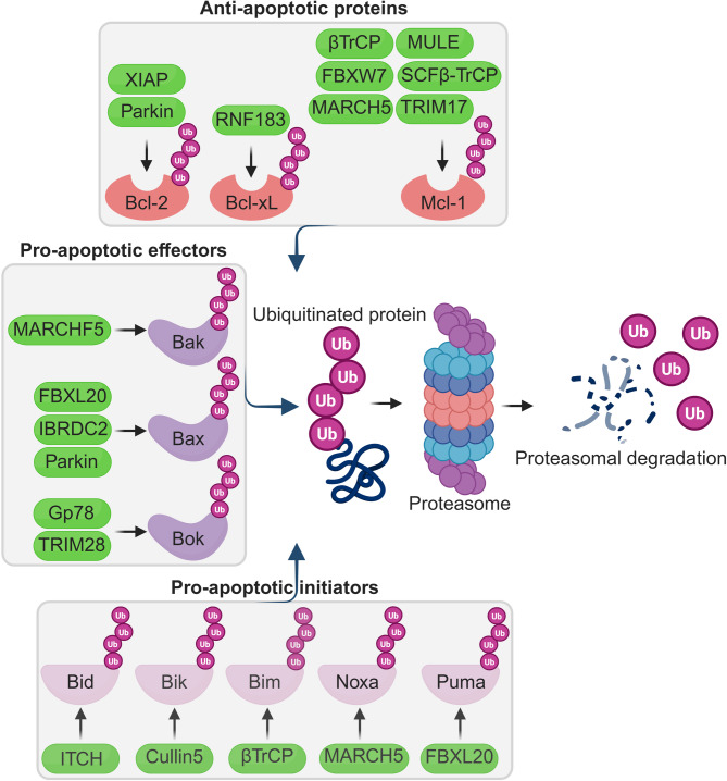
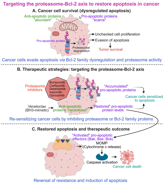
The B-cell lymphoma-2 (Bcl-2) family proteins, key regulators of apoptosis, are frequently dysregulated in cancer, tipping the balance of cell survival and apoptosis in favor of survival. The ubiquitin-proteasome system (UPS) is a critical cellular machinery that controls the Bcl-2 levels through regulation of protein stability. This review delves into the intricate interplay between the proteasome and Bcl-2 family members, exploring how proteasome-mediated degradation impacts cell survival and proliferation to influence cancer progression. We discuss the therapeutic potential of targeting the proteasome-Bcl-2 axis, including the use of proteasome inhibitors as anticancer agents. We examine their mechanisms of action, clinical efficacy, and limitations while exploring emerging strategies to overcome these challenges.

Keywords: Apoptosis; Bcl-2 family proteins; Cancer; Proteasome inhibitors; Therapeutic potential; Ubiquitin-proteasome system (UPS); Ubiquitination.


Conflict of interest statement

Declarations. Ethics approval and consent to participate: Not applicable. Consent for publication: Not applicable. Competing interests: The authors declare no competing interests.

Figures

Fig. 1
Fig. 1
The intrinsic mitochondrial pathway of apoptosis: central role of Bcl-2 family proteins in stress-induced cell death. Cytotoxic stress signals such as DNA damage, nutrient deprivation, or reactive oxygen species initiate intracellular stress responses that activate BH3-only pro-apoptotic initiators (Bad, Bid, Bik, Bim, Noxa, Puma). These proteins either directly activate pro-apoptotic effectors (Bak, Bax, Bok) or indirectly promote their activation by neutralizing anti-apoptotic members (Bcl-2, Bcl-xL, Mcl-1). Once activated, Bak/Bax/Bok oligomerize at the mitochondrial outer membrane, causing mitochondrial outer membrane permeabilization (MOMP) and subsequent release of cytochrome c into the cytoplasm. This leads to apoptosome formation, caspase activation, and apoptosis. Arrows denote activation, while blunt-ended lines indicate inhibition. Red text indicates BH3-only pro-apoptotic initiators, orange indicates pro-apoptotic effectors, and green indicates anti-apoptotic proteins. Overall, this figure illustrates how Bcl-2 family members integrate cytotoxic stress signals to regulate mitochondrial-mediated apoptosis
Fig. 2
Fig. 2
Ubiquitin-proteasome system (UPS)-mediated regulation of Bcl-2 family proteins. This figure illustrates the role of E3 ubiquitin ligases in targeting Bcl-2 family proteins for ubiquitination and subsequent proteasomal degradation. Central to the figure is the ubiquitin-proteasome system (UPS), where polyubiquitinated proteins are recognized and degraded by the 26 S proteasome. Proteins marked with multiple “Ub” (ubiquitin) icons are designated for degradation. Top panel: Anti-apoptotic Bcl-2 family proteins (Bcl-2, Bcl-xL, and Mcl-1) are regulated by specific E3 ligases (green ovals) which promote their ubiquitination. Left panel: Pro-apoptotic effector proteins (Bak, Bax, and Bok) are similarly targeted by specific E3 ligases (green ovals) to modulate apoptosis. Bottom panel: BH3-only pro-apoptotic initiator proteins (Bid, Bik, Bim, Noxa, Puma) are shown to be ubiquitinated by specific E3 ligases (green ovals). The central flow in the figure depicts the general UPS process, E3 ligases catalyze the attachment of ubiquitin to substrates, leading to recognition and degradation by the proteasome. Ubiquitinated proteins (tagged with purple Ub chains) enter the proteasome, are degraded, and free ubiquitin molecules are recycled. Green ovals indicate E3 ubiquitin ligases; arrows denote direction of ubiquitination or degradation. Shapes and colors of Bcl-2 family proteins represent their functional class: red (anti-apoptotic), purple (pro-apoptotic effectors), and pink (BH3-only proteins). Ub = Ubiquitin
Fig. 3
Fig. 3
Proteasome inhibition restores apoptosis in cancer by preventing degradation of pro-apoptotic proteins. This figure illustrates how the ubiquitin-proteasome system (UPS) contributes to tumor cell survival by targeting pro-apoptotic proteins for degradation. In cancer cells, anti-apoptotic proteins are frequently overexpressed, while pro-apoptotic proteins are downregulated or degraded via the UPS, promoting tumor growth. Top panel: Cancer cells upregulate anti-apoptotic proteins and suppress pro-apoptotic proteins, enabling survival and uncontrolled proliferation. Center panel: Pro-apoptotic proteins are ubiquitinated and targeted for proteasomal degradation. The figure depicts the 26 S proteasome recognizing polyubiquitinated proteins and degrading them into peptides, thereby suppressing apoptosis. Bottom panel: FDA-approved proteasome inhibitors (Bortezomib, Carfilzomib, Delanzomib, Ixazomib, Oprozomib) block proteasomal activity, leading to accumulation of pro-apoptotic proteins and restoration of apoptotic signaling in tumor cells. By preventing the degradation of key pro-apoptotic factors, proteasome inhibitors re-activate apoptosis, offering a therapeutic strategy against proteasome-addicted tumors. Purple circles labeled “Ub” indicate ubiquitin. Dashed arrows show degradation; blocked lines indicate inhibition
Fig. 4
Fig. 4
Targeting the proteasome-Bcl-2 axis to restore apoptosis in cancer. This summary illustration depicts the interplay between the ubiquitin-proteasome system (UPS), Bcl-2 family proteins, and therapeutic strategies aimed at reinstating apoptosis in cancer cells. (A) Cancer cell survival (dysregulated apoptosis): In cancer, apoptotic resistance is driven by overexpression of anti-apoptotic Bcl-2 family members (e.g., Bcl-2, Bcl-xL, Mcl-1, shown in green) and proteasomal degradation of pro-apoptotic proteins (e.g., Bax, Bak, Bok, and BH3-only proteins such as Bim, Noxa, Puma, etc. shown in purple). Active proteasomes degrade key pro-apoptotic regulators`, resulting in their reduced cellular levels and enabling evasion of mitochondrial outer membrane permeabilization (MOMP), caspase activation, and apoptosis. This imbalance promotes unchecked proliferation and tumor survival. (B) Therapeutic strategies: targeting the proteasome–Bcl-2 axis: The top branch illustrates proteasome inhibition using small molecules (e.g., Bortezomib, Carfilzomib, Delanzomib, Ixazomib, Oprozomib), which blocks degradation of pro-apoptotic proteins, leading to their accumulation and restored apoptotic potential. The bottom branch depicts BH3 mimetics (e.g., Venetoclax), which bind to and neutralize anti-apoptotic proteins, thereby releasing sequestered pro-apoptotic effectors. Both strategies restore the pro-apoptotic/anti-apoptotic balance, sensitizing cancer cells to apoptosis. (C) Restored apoptosis and therapeutic outcome: Rebalanced Bcl-2 signaling enables activation of pro-apoptotic effectors (Bak, Bax, Bok), MOMP, release of cytochrome c, and activation of the apoptosome and caspase cascade. This culminates in the execution of apoptosis, characterized by morphological changes such as membrane blebbing and nuclear fragmentation, ultimately leading to cancer cell death, tumor regression, and improved therapeutic outcomes. Color coding: Anti-apoptotic proteins (green), pro-apoptotic proteins (purple), proteasome inhibitors (red). Arrows indicate activation or consequence; dashed lines represent inhibitory effects

References

    1. Voss AK, Strasser A. The essentials of developmental apoptosis. F1000Res 2020;9:148. - PMC - PubMed
    1. Fogarty CE, Bergmann A. Killers creating new life: caspases drive apoptosis-induced proliferation in tissue repair and disease. Cell Death Differ. 2017;24:1390–400. - PMC - PubMed
    1. Hanggi K, Ruffell B. Cell death, therapeutics, and the immune response in cancer. Trends Cancer. 2023;9:381–96. - PMC - PubMed
    1. Xu J, Dong X, Huang DCS, Xu P, Zhao Q, Chen B. Current advances and future strategies for BCL-2 inhibitors: potent weapons against cancers. Cancers (Basel) 2023;15:4957. - PMC - PubMed
    1. Kale J, Osterlund EJ, Andrews DW. BCL-2 family proteins: changing partners in the dance towards death. Cell Death Differ. 2018;25:65–80. - PMC - PubMed

MeSH terms

Substances

LinkOut - more resources