

High-affinity transferrin receptor binding improves brain delivery of bispecific antibodies at tracer dose
- PMID: 40842024
- PMCID: PMC12369151
- DOI: 10.1186/s12987-025-00693-2
High-affinity transferrin receptor binding improves brain delivery of bispecific antibodies at tracer dose
Abstract
Background: Transferrin receptor (TfR)-mediated transcytosis is a well-established method for delivering biologic therapeutics and diagnostics to the brain. Although moderate affinity towards TfR is beneficial for TfR-mediated brain delivery at therapeutic doses, emerging evidence has indicated that high TfR affinity may be more beneficial at tracer doses. With the development of antibody-based PET radioligands for neurodegenerative diseases, such as Alzheimer's disease, understanding the pharmacokinetics of TfR-binders at tracer dose is essential. Thus, this study aimed to evaluate the effect of TfR affinity on brain uptake at a tracer dose in both wild-type (WT) and amyloid-beta (Aβ) pathology presenting mice and to demonstrate the usability of TfR-mediated brain delivery of immunoPET diagnostic radioligands to visualize intrabrain Aβ pathology in vivo.
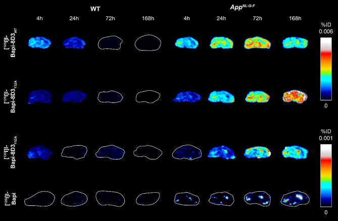
Methods: Three different affinity variants of anti-mouse TfR-binding antibody 8D3, engineered by alanine point mutations, were selected. Bispecific antibodies were designed with knob-into-hole technology with one arm targeting TfR (8D3) and the other arm targeting human Aβ (bapineuzumab). Antibody affinities were measured in an in vitro cell assay. In vivo pharmacokinetic analyses of radioiodinated bispecific antibodies and bapineuzumab in brain, blood and peripheral organs were performed over 7 days post-injection in WT mice and a model of Aβ pathology (AppNL-G-F). The strongest TfR affinity bispecific antibody was also evaluated as a positron emission tomography (PET) radioligand for detecting Aβ pathology in WT and AppNL-G-F mice.
Results: The three bispecific antibodies bound to TfR with affinities of 10 nM, 20 nM and 240 nM. Independent of genotype, stronger TfR-affinity resulted in higher initial brain uptake. The two higher-affinity bispecific antibodies behaved similarly and differentiated between WT and AppNL-G-F mice earlier than the lowest affinity variant. Finally, the 10 nM bispecific antibody was able to clearly differentiate between WT and AppNL-G-F mice when used as a PET radioligand.
Conclusion: This study supports the hypothesis that stronger TfR affinity enhances brain uptake at a tracer dose. With the more effective detection of Aβ pathology, stronger TfR affinity is a crucial design feature for future bispecific immunoPET radioligands for intrabrain targets via TfR-mediated transcytosis.
Keywords: Affinity; Alzheimer’s disease; Amyloid-β; Positron emission tomography (PET); Transferrin receptor (TfR)-mediated transcytosis.
© 2025. The Author(s).
Conflict of interest statement
Declarations. Ethics approval: All animal experiments described in this study were approved by the Uppsala County Animal Ethics board (5.8.18–20401/2020), following the rules and regulations of the Swedish Animal Welfare Agency and complied with the European Communities Council Directive of 22 September 2010 (2010/63/EU). Consent for publication: Not applicable. Competing interests: GB, SSi, LS and KGA are employees of BioArctic AB, Sweden. The other authors declare that they have no competing interests.
Figures


Similar articles
-
Fluorine-18 ImmunoPET Imaging of Antibody Brain Kinetics and Amyloid-Beta Pathology.ACS Pharmacol Transl Sci. 2025 Jul 11;8(8):2804-2813. doi: 10.1021/acsptsci.5c00359. eCollection 2025 Aug 8. ACS Pharmacol Transl Sci. 2025. PMID: 40810143
-
PET imaging of TREM2 in amyloid-beta induced neuroinflammation.Eur J Nucl Med Mol Imaging. 2025 Sep;52(11):4320-4333. doi: 10.1007/s00259-025-07358-0. Epub 2025 May 28. Eur J Nucl Med Mol Imaging. 2025. PMID: 40434494 Free PMC article.
-
Serotransferrin enhances transferrin receptor-mediated brain uptake of antibodies.Drug Deliv Transl Res. 2025 Sep;15(9):3321-3337. doi: 10.1007/s13346-025-01811-1. Epub 2025 Feb 19. Drug Deliv Transl Res. 2025. PMID: 39971861 Free PMC article.
-
Bispecific brain-penetrant antibodies for treatment of Alzheimer's disease.J Prev Alzheimers Dis. 2025 Sep;12(8):100214. doi: 10.1016/j.tjpad.2025.100214. Epub 2025 May 26. J Prev Alzheimers Dis. 2025. PMID: 40425446 Review.
-
18F PET with flutemetamol for the early diagnosis of Alzheimer's disease dementia and other dementias in people with mild cognitive impairment (MCI).Cochrane Database Syst Rev. 2017 Nov 22;11(11):CD012884. doi: 10.1002/14651858.CD012884. Cochrane Database Syst Rev. 2017. PMID: 29164602 Free PMC article.
References
-
- Mukherjee AG, Wanjari UR, Gopalakrishnan AV, et al. Evolving strategies and application of proteins and peptide therapeutics in cancer treatment. Biomedicine and Pharmacotherapy. Volume 163. Elsevier Masson s.r.l.; 2023. - PubMed
-
- Zhou J, Deng H, Xiao H. Research and development of antibody-based novel anti-cancer drugs: an overview. Intelligent Pharmacy. Volume 1. KeAi Publishing Communications Ltd.; 2023.
-
- Magnusson K, Sehlin D, Syvänen S, et al. Specific uptake of an amyloid-β protofibril-binding antibody-tracer in AβPP Transgenic mouse brain. J Alzheimer’s Disease. 2013;37:29–40. - PubMed
MeSH terms
Substances
LinkOut - more resources
Full Text Sources
