. 2025 Aug;29(16):e70741.
doi: 10.1111/jcmm.70741.

Sirt4 Deficiency Promotes Cardiomyocyte Proliferation and Cardiac Repair

Affiliations

Sirt4 Deficiency Promotes Cardiomyocyte Proliferation and Cardiac Repair

Weijing Liu et al. J Cell Mol Med. 2025 Aug.

Abstract

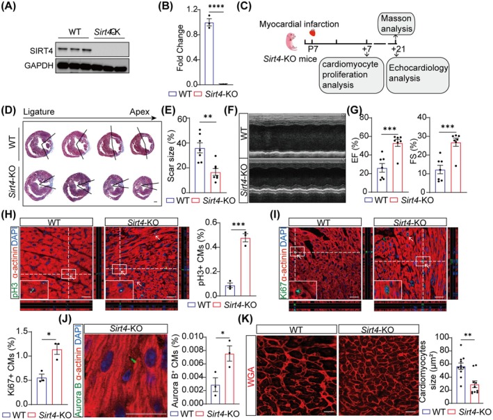
The mammalian heart exhibits transient but remarkable regenerative capacity during the early postnatal period, after which most cardiomyocytes exit the cell cycle. While the sirtuin family is well-established as regulators of cell cycle progression, its specific role in cardiomyocyte proliferation and cardiac regeneration remains unclear. In this study, we found that Sirt4 expression increased during postnatal heart development. Adenovirus-mediated Sirt4 overexpression in vitro inhibited cardiomyocyte proliferation by inducing oxidative DNA damage. Moreover, cardiomyocyte-specific Sirt4 overexpression in vivo suppressed cardiomyocyte proliferation and impaired neonatal heart regeneration. Using Sirt4-knockout mice, we found that Sirt4 deficiency promoted cardiomyocyte proliferation and extended the heart regeneration window. Furthermore, Sirt4 deficiency improved cardiac function and reduced myocardial fibrosis after ischaemia-reperfusion injury in adult mice. These findings establish Sirt4 as a critical regulator of cardiomyocyte proliferation and cardiac repair, suggesting that targeted Sirt4 inhibition may represent a promising therapeutic strategy for ischaemic heart diseases.

Keywords: Sirt4; cardiomyocyte proliferation; heart regeneration; oxidative DNA damage.


Conflict of interest statement

The authors declare no conflicts of interest.

Figures

FIGURE 1
FIGURE 1
Sirt4 overexpression inhibits cardiomyocyte proliferation in vitro. (A, B) Boxplot showing RNA expression of Sirtuins genes in mouse heart samples during heart development (A) or subjected to myocardial infarction (MI) (B) based on RNA‐sequencing data (n = 3 biological replicates). P0, postnatal Day 0; P60, postnatal Day 60; dps, day post sham; dpi, day post infarction. (C) Schematic representation of the experiment design. Neonatal mouse cardiomyocytes isolated from P1 mice (P1‐NMCMs) were infected with Ad‐Sirt4 and Ad‐Sirt5 for 48 h, with Ad‐NC treatment was the controls. WT, wild type; Ad‐NC, adenovirus (Ad) harbouring a vector control; Ad‐Sirt4, adenovirus (Ad) harbouring Sirt4; Ad‐Sirt5, adenovirus (Ad) harbouring Sirt5. (D, E) Quantitative real‐time polymerase chain reaction (qRT‐PCR) analysis of Sirt4 and Sirt5 expression in P1 neonatal mice cardiomyocytes (P1‐NMCMs) infected with Ad‐NC and Ad‐Sirt4, Ad‐Sirt5 for 48 h (n = 3 biological replicates). (F) Western blot analysis of SIRT4 and SIRT5 expression in P1‐NMCMs infected with Ad‐NC and Ad‐Sirt4, or Ad‐Sirt5 for 48 h (n = 3 biological replicates). (G–I) Immunofluorescence analysis of the proliferation of P1‐NMCMs infected with Ad‐NC and Ad‐Sirt4 for 48 h (N = 8 biological replicates for pH 3+ and Ki67+ CMs, n = 3 biological replicates for Aurora B+ CMs). White arrows indicate pH 3+, Ki67+ or Aurora B+ CMs. Scale bars, 20 μm. (J) Immunofluorescence analysis of the proliferation of P1‐NMCMs infected with Ad‐NC and Ad‐Sirt5 for 48 h (n = 6 biological replicates). White arrows indicate pH 3+ CMs. Scale bars, 20 μm. Data are mean ± SEM; *p < 0.05, **p < 0.01, ***p < 0.001, ****p < 0.0001; ns, not significance; unpaired two‐tailed t‐tests (A, B, D–J).
FIGURE 2
FIGURE 2
Ectopically expressed Sirt4 impairs heart regeneration in neonatal mice. (A, B) Western blot analysis (A) and quantification (B) of SIRT4 expression in N‐Tg and Sirt4‐Tg mice at 7 dpr (n = 3 biological replicates). N‐Tg, negative transgenic; Sirt4‐Tg, Sirt4 transgenic. (C) Schematic representation of the experiment design. Apical resection was performed on postnatal Day 1 (P1) mice. Hearts were harvested for cardiomyocytes (CMs) proliferation analysis at 7‐day post resection (dpr) and for myocardial fibrosis and echocardiology analysis at 21 dpr. (D, E) Representative Masson trichrome staining of heart sections and the quantification of fibrosis area proportion of N‐Tg and Sirt4‐Tg mice at 21 dpr (n = 9 mice). Scale bars, 400 μm. (F) Representative images of M‐Mode echocardiographic assessment. (G) The ejection fraction (EF) and fractional shortening (FS) of the left ventricle in N‐Tg and Sirt4‐Tg mice at 21 dpr (n = 9 mice). (H–J) Immunofluorescence analysis of CM proliferation in heart sections at 7 dpr (n = 3 mice). White arrows indicate pH 3+ or Ki67+, Aurora B+ CMs. Scale bars, 20 μm. (K) Immunofluorescence images and quantification of mononucleated, binucleated, and multinucleated CMs isolated at 14 dpr from N‐Tg and Sirt4‐Tg mice (n = 4 mice). Scale bars, 20 μm. Data are mean ± SEM; *p < 0.05, **p < 0.01, ****p < 0.0001; ns, not significance; unpaired two‐tailed t‐tests (B, E, G–J); one‐way ANOVA (K).
FIGURE 3
FIGURE 3
Sirt4 overexpression activates oxidative DNA damage response in cardiomyocytes. (A) Immunofluorescence analysis of Sirt4 location in primary cardiomyocytes isolated from P1 mice (P1‐NMCMs) infected with Ad‐Sirt4 for 48 h. Mito, mitochondria. Scale bars, 10 μm. (B) Quantitative real‐time polymerase chain reaction (qRT‐PCR) analysis of mitochondrial DNA content using NADH dehydrogenase subunit 1 (mtND1), encoded by mitochondrial DNA in P1‐NMCMs infected with Ad‐NC and Ad‐Sirt4 for 48 h (n = 6 biological replicates). Ad‐NC, adenovirus (Ad) harbouring a vector control; Ad‐Sirt4, adenovirus (Ad) harbouring Sirt4. (C) Immunofluorescence analysis of mitochondria in P1‐NMCMs infected with Ad‐Sirt4 for 48 h. Mito, mitochondria. Scale bars, 10 μm. (D) Quantification analysis of the number of individual mitochondria in P1‐NMCMs infected with Ad‐NC and Ad‐Sirt4 for 48 h (n = 15 biological replicates for Ad‐NC and n = 19 biological replicates for Ad‐Sirt4). (E) Quantification analysis of the number of network mitochondria in P1‐NMCMs infected with Ad‐NC and Ad‐Sirt4 for 48 h (n = 18 biological replicates for Ad‐NC and n = 19 biological replicates for Ad‐Sirt4). (F) Quantification analysis of the number of individual mitochondria compared to network mitochondria in P1‐NMCMs infected with Ad‐NC and Ad‐Sirt4 for 48 h (n = 18 biological replicates for Ad‐NC and n = 18 biological replicates for Ad‐Sirt4). (G) Transmission electron microscopy images of P1‐NMCMs infected with Ad‐NC and Ad‐Sirt4 for 48 h. Scale bars, 0.5 μm. M, Mitochondria; S, Sarcomere. (H, I) Quantification analysis of transmission electron microscopy images in G. (J) Flow cytometry analysis of reactive oxygen species (ROS) level in P1‐NMCMs infected with Ad‐Sirt4 for 48 h (n = 4 biological replicates for Ad‐NC and n = 5 biological replicates for Ad‐Sirt4). (K, L) Immunofluorescence analysis of oxidative DNA damage (indicated by oxidatively modified base 8OHG) and phosphorylated ATM (pATM) expression in P1‐NMCMs infected with Ad‐Sirt4 for 48 h (n = 6 biological replicates). Scale bars, 20 μm. Data are mean ± SEM; *p < 0.05, **p < 0.01, ****p < 0.0001; unpaired two‐tailed t‐tests (B, D–F, H–L).
FIGURE 4
FIGURE 4
Sirt4 deficiency induces juvenile heart regeneration. (A, B) Western blot analysis (A) and quantification (B) of SIRT4 expression in P7 WT and Sirt4‐KO mice (n = 3 biological replicates). WT, wild type; Sirt4‐KO, Sirt4 knockout. (C) Schematic representation of the experiment design. Myocardial infarction (MI) was performed on postnatal Day 7 (P7) mice. Hearts were harvested for gene expression and cardiomyocytes (CMs) proliferation analysis at 7‐day post infarction (dpi); and for myocardial fibrosis and echocardiology analysis at 21 dpr. (D, E) Representative Masson trichrome staining of heart cross‐sections and the quantification of fibrosis area proportion of WT and Sirt4‐KO mice at 21 dpi (n = 7 mice). Scale bars, 400 μm. (F) Representative images of M‐Mode echocardiographic assessment. (G) The ejection fraction (EF) and fractional shortening (FS) of the left ventricle in WT and Sirt4‐KO mice at 21 dpi (n = 7 mice). (H–J) Immunofluorescence analysis of CM proliferation in heart cross‐sections at 7 dpi (n = 3 mice). White arrows indicate pH 3+ or Ki67+, Aurora B+ CMs. Scale bars, 20 μm. (K) Immunofluorescence analysis of CM size in heart cross‐sections at 7 dpi (n = 3 mice). Scale bars, 20 μm. Data are mean ± SEM; *p < 0.05, **p < 0.01, ***p < 0.001, ****p < 0.0001; unpaired two‐tailed t‐tests (B, E, G–K).
FIGURE 5
FIGURE 5
Sirt4 deficiency promotes heart regeneration in adult mice. (A) Schematic representation of the experiment design. Ischemia–reperfusion (I‐R) was performed in 8 weeks mice. The hearts were harvested for cardiomyocyte (CM) proliferation analysis at 7‐day post I‐R; for scar size analysis with Masson trichrome staining at 28‐day post I‐R. Echocardiology analysis was performed at before and 4, 7 and 28 days post I‐R. WT, wild type; Sirt4‐KO, Sirt4 knockout. (B) Representative Masson's trichrome staining of heart cross‐sections and the quantification of fibrosis area proportion of WT and Sirt4‐KO mice at 28‐day post I‐R (n = 7 mice). Scale bars, 500 μm. WT, wild type. (C) Representative images of M‐Mode echocardiographic assessment. (D, E) Ejection fraction (EF) and fractional shortening (FS) of the left ventricle in WT and Sirt4‐KO mice before and 4, 7 and 28 days post I‐R (n = 7 mice). (F–H) Immunofluorescence analysis of CM proliferation in heart cross‐sections at 7‐day post I‐R (n = 3 mice). White arrows indicate pH 3+ or Ki67+, Aurora B+ CMs. Scale bars, 20 μm. Data are mean ± SEM; *p < 0.05, **p < 0.01, ***p < 0.001; unpaired two‐tailed t‐tests (B, F–H); two‐way ANOVA (D, E).

Similar articles

References

    1. Doiron J. E. and Lefer D. J., “Leveraging Adipocyte‐Cardiomyocyte Signaling to Treat Ischemic Heart Failure,” Circulation Research 131 (2022): 165–167. - PubMed
    1. Zhu C., Yuan T., and Krishnan J., “Targeting Cardiomyocyte Cell Cycle Regulation in Heart Failure,” Basic Research in Cardiology 119 (2024): 349–369. - PMC - PubMed
    1. Xing M., Jiang Y., Bi W., et al., “Strontium Ions Protect Hearts Against Myocardial Ischemia/Reperfusion Injury,” Science Advances 7 (2021): eabe0726. - PMC - PubMed
    1. Laflamme M. A. and Murry C. E., “Heart Regeneration,” Nature 473 (2011): 326–335. - PMC - PubMed
    1. Tzahor E. and Poss K. D., “Cardiac Regeneration Strategies: Staying Young at Heart,” Science 356 (2017): 1035–1039. - PMC - PubMed

MeSH terms

LinkOut - more resources