. 2025 Nov 5;33(11):5489-5504.
doi: 10.1016/j.ymthe.2025.08.023. Epub 2025 Aug 20.

Exploiting viral infection/vaccination to focus high-affinity T cell populations into tumors using oncolytic viro-immunotherapy

Affiliations

Exploiting viral infection/vaccination to focus high-affinity T cell populations into tumors using oncolytic viro-immunotherapy

Alexa Veliz Rios et al. Mol Ther. .

Abstract

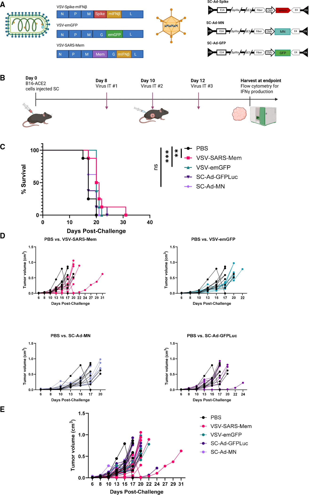
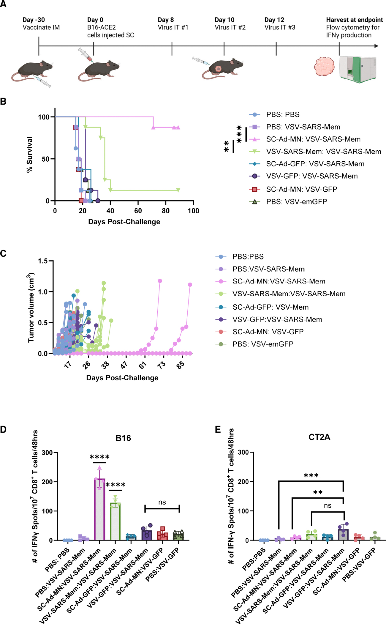
Immune tolerance restricts the number of T cells with significant affinity for self-tumor-associated antigens (TAAs), thereby limiting successful cancer immunotherapy through an inability to generate populations of high-affinity anti-tumor T cells. In contrast, viral infection/vaccination primes and expands high-affinity effector and memory T cells against viral antigens. We show here that it is possible to exploit population-wide preexisting, anti-viral memory recall responses against SARS-CoV-2 antigens to focus a high-affinity, immunodominant T cell response into tumors by oncolytic virus (OV)-mediated or chimeric antigen receptor (CAR)-mediated delivery of viral antigens that are not themselves related to TAAs. Heterologous prime and OV/boost led to CD8+ T cell-dependent tumor cures using either SARS-CoV-2 Mem or Spike (S) proteins as vaccinating/tumor-focusing T cell targets, associated with epitope spreading against TAAs. We also show that CAR-T cells carry SARS-CoV-2 antigen-expressing vectors systemically to tumors even in pre-immune mice. Finally, S-specific CAR-T cells could be boosted in vivo with S protein vaccines to enhance anti-tumor activity and persistence. Thus, where high affinity anti-tumor T cells are not available, boosting preexisting infection- or vaccination-induced T cell populations within tumors using OV-mediated immunogen delivery provides a therapeutically valuable alternative.

Keywords: SARS-CoV-2; cancer immunotherapy; oncolytic viruses; single cycle adenovirus; tumor antigens; vaccines; vesicular stomatitis virus; virus T cell memory.


Conflict of interest statement

Declaration of interests The authors declare no competing interests.

Figures

Figure 1.
Figure 1.. VSV-SARS-Mem has modest activity as an oncolytic in the B16 model
(A) Schematic of adenoviral and VSV vectors expressing GFP, Spike (S), or the membrane (Mem) protein of SARS-CoV-2. (B) C57BL6 mice bearing B16-human ACE2 tumors received three intratumoral (IT) injections with VSV (108 PFU/injection) or adenovirus (Ad) (1010 vp/injection). Survival with time is shown (C) **p = 0.0058 for PBS against VSV-SARS-Mem; ***p = 0.0019 for PBS against VSV-emerald(em)GFP; ns, statistically non-significant; log rank test with Grehan-Breslow-Wilcoxon test. (D and E) (D) Individual and (E) combined tumor volumes across different treatment groups.
Figure 2.
Figure 2.. Recruitment of a SARS-CoV-2 Mem protein recall response by VSV-Mem OV cures tumors
(A) C57BL6 mice were intramuscularly (IM) vaccinated 30 days prior to B16-ACE2 tumor inoculation with either VSV (108 PFU) or Ad (1010 vp). Tumors were treated IT 8, 10, and 12 days after tumor seeding, with VSV expressing SARS-Mem or emGFP (108 PFU/injection). (B) Survival with time is shown; **p ≤ 0.01, ***p ≤ 0.001; log rank test with Grehan-Breslow-Wilcoxon test. (C) Tumor volumes across different treatment groups. The majority of mice treated with VSV-Mem: VSV-Mem reached endpoint by day 40. The only mouse surviving developed a small tumor 26 days post-tumor cell challenge, which completely regressed by day 38. (D and E) IFN-γ ELISpot from CD8+ cells isolated from splenocytes of different treatment groups re-stimulated in vitro with live B16 melanoma (D) or irrelevant CT2A glioma (E) cells as targets (E:T ratio 10:1). Error bars denote SD. **p ≤ 0.01, ***p ≤ 0.001; ****p ≤ 0.0001; ns, statistically non-significant; one-way ANOVA.
Figure 3.
Figure 3.. IT focusing of anti-SARS-Mem immunity for tumor therapy depends upon CD8+ and CD4+ T cells
(A) C57BL6 mice were IM vaccinated 30 days prior to B16-ACE2 tumor inoculation with SC-Ad (1010 vp). At 8, 10, 12, 15, 17, and 19 days after tumor seeding, tumors were treated IT with VSV expressing Mem or emGFP (108 PFU). Depleting antibodies for CD4+, CD8+ and natural killer (NK) cells were injected on days 8, 9, 10, 15, 16, and 17. (B) Survival with time is shown; *p ≤ 0.05, ****p ≤ 0.0001; ns, statistically non-significant; log rank test with Grehan-Breslow-Wilcoxon test. (C) Tumor volumes across diffesrent treatment groups.
Figure 4.
Figure 4.. S-immune mice are effectively treated with S-expressing virotherapy
(A) ACE2-expressing tumor cells infected with VSV-S generate large multi-nucleated syncytia (one example highlighted with red arrows) 48 h post-infection. (B) C57BL6 mice were vaccinated IM 30 days prior to seeding of B16-ACE2 tumors with SC-Ad (1010vp) expressing either Mem or S. At 8, 10, 12, 17, 19, and 21 days later tumors were treated IT with either VSV (108 PFU) or SC-Ad (1010 vp) expressing Mem or S. (C) Survival with time is shown; ***p ≤ 0.001, ****p ≤ 0.0001; ns, statistically non-significant; log rank test with Grehan-Breslow-Wilcoxon test. (D) Tumor volumes across different treatment groups. (E) IFN-γ ELISpot from CD8+ cells isolated from splenocytes of different treatment groups re-stimulated in vitro with live B16 melanoma (E) or irrelevant CT2A glioma (F) cells as targets (E:T ratio 10:1). Error bars denote SD. ****p ≤ 0.0001; ns, statistically non-significant; one-way ANOVA.
Figure 5.
Figure 5.. CAR-mediated delivery of recall viral antigens allows for systemic therapy
(A) B16-EGFRvIII tumors were seeded in C57Bl/6 k18-hACE2 mice (n = 7–8/group). On days 7, 9, and 11 mice were either left unimmunized or were immunized with an S-derived peptide library (1 μg/mouse/injection). On days 14, 16, and 18, groups were treated with unloaded anti-EGFRvIII CAR-T cells (107 CAR-T cells/injection), with CAR-T cells loaded ex vivo with SC-Ad-GFP or with CAR-T cells loaded ex vivo with SC-Ad-S (107 CAR-T cells, loaded ex vivo at 4°C, MOI 1.0). (B) Survival with time is shown; ***p ≤ 0.001; ****p ≤ 0.0001; log rank test with Grehan-Breslow-Wilcoxon test. (C and D) IFN-γ ELISpot from splenocytes isolated from different treatment groups re-stimulated in vitro with live B16 melanoma (C) or CAR target antigen expressing CT2A-EGFRvIII glioma (D) cells as targets (E:T ratio 1:1). Error bars denote SD. ***p ≤ 0.001; ****p ≤ 0.0001; ns, statistically non-significant; one-way ANOVA. (E and F) IFN-γ ELISpot from splenocytes recovered from different treatment groups re-stimulated in vitro with S-specific peptide library or irrelevant ovalbumin-derived SIINFEKL peptide; Error bars denote SD. *p ≤ 0.05, **p ≤ 0.001, ****p ≤ 0.0001; ns, statistically non-significant; one-way ANOVA.
Figure 6.
Figure 6.. CAR-T cells from pre-immune individuals can be boosted in vivo for improved therapy
(A) B16-EGFRvIII tumors were seeded in C57Bl/6 k18-hACE2 mice (n = 7–8/group). On days 7, 9, and 11 mice (n = 7–8 mice/group) were treated with anti-EGFRvIII CAR T cells (107/injection), which were isolated from either naive C57Bl/6 donors or from C57Bl/6 donor mice vaccinated 30 days previously with the Spike peptide library (1 μg Peptivator library/vaccine with poly(I:C) adjuvant) (CARimmune). On days 14, 16, and 18, mice were either boosted with the irrelevant ovalbumin-derived SIINFEKL peptide or with the Spike-derived peptide library (1 μg/boost). (B) Survival with time is shown; ***p ≤ 0.001; log rank test with Grehan-Breslow-Wilcoxon test. (C) Mice that rejected their tumors following treatment with S-boosted CARimmune in (B) (n = 8) and naive C57Bl/6 k18-hACE2 mice were challenged IN with VSV-S (5 ×10⁵ PFU/nostril, which is lethal to unprotected C57Bl/6 k18-hACE2 mice) as a surrogate model of SARS-CoV-2 infection. **p ≤ 0.001, log rank test with Grehan-Breslow-Wilcoxon test. Survival with time is shown.

References

    1. Topalian SL, Forde PM, Emens LA, Yarchoan M, Smith KN, and Pardoll DM (2023). Neoadjuvant immune checkpoint blockade: A window of opportunity to advance cancer immunotherapy. Cancer Cell 41, 1551–1566. 10.1016/j.ccell.2023.07.011. - DOI - PMC - PubMed
    1. Morad G, Helmink BA, Sharma P, and Wargo JA (2021). Hallmarks of response, resistance, and toxicity to immune checkpoint blockade. Cell 184, 5309–5337. 10.1016/j.cell.2021.09.020. - DOI - PMC - PubMed
    1. Fenis A, Demaria O, Gauthier L, Vivier E, and Narni-Mancinelli E (2024). New immune cell engagers for cancer immunotherapy. Nat. Rev. Immunol. 24, 471–486. 10.1038/s41577-023-00982-7. - DOI - PubMed
    1. Katsikis PD, Ishii KJ, and Schliehe C (2024). Challenges in developing personalized neoantigen cancer vaccines. Nat. Rev. Immunol. 24, 213–227. 10.1038/s41577-023-00937-y. - DOI - PMC - PubMed
    1. Rosenberg SA, and Restifo NP (2015). Adoptive cell transfer as personalized immunotherapy for human cancer. Science 348, 62–68. 10.1126/science.aaa4967. - DOI - PMC - PubMed

MeSH terms