. 2025 Dec;16(1):2548625.
doi: 10.1080/21505594.2025.2548625. Epub 2025 Aug 21.

PRRSV evades innate immune cGAS-STING antiviral function via its Nsp5 to deter STING translocation and activation

Affiliations

PRRSV evades innate immune cGAS-STING antiviral function via its Nsp5 to deter STING translocation and activation

Yulin Xu et al. Virulence. 2025 Dec.

Abstract

Porcine Reproductive and Respiratory Syndrome Virus (PRRSV) is an important pathogen that seriously endangers pig breeding, causing significant economic losses to the global swine industry. Our previous study found that the DNA sensing innate cGAS-STING signaling pathway plays an important role in inducing interferon (IFN) upon PRRSV infection and inhibition of PRRSV replication. However, the mechanism underlying immune evasion by PRRSV remains unclear. In the current study, we found that PRRSV non-structural protein 5 (Nsp5) strongly inhibits the cGAS-STING-IFN antiviral response. Furthermore, we found that Nsp5 interacts with STING, blocking STING transport from the ER to the Golgi apparatus and interfering with STING recruitment of TBK1/IKKε/IRF3. Finally, we demonstrated that the Nsp5 36-47 and 58-67 amino acid regions are critical for inhibiting STING activity and PRRSV replication. This study describes a novel mechanism by which PRRSV suppresses the host innate antiviral response and has implications for our understanding of PRRSV pathogenesis.

Keywords: Nsp5; PRRSV; STING; immune evasion.


Conflict of interest statement

No potential conflict of interest was reported by the author(s).

Figures

Figure 1.
Figure 1.
PRRSV proteins Nsp3, Nsp5 and E interact with STING. (A-C) HEK-293T cells were co-transfected pSTING-HA together with the indicated plasmids encoding GFP-tagged PRRSV viral proteins for 24 h. The cells were harvested for immunoprecipitation using rabbit anti-HA antibody, and IP samples and cell lysate inputs were subjected to Western blotting by using mouse anti-HA antibody and mouse anti-GFP antibody. (D) HEK-293T cells were co-transfected pSTING-HA together with the plasmids encoding GFP-tagged PRRSV viral proteins (Nsp3, Nsp5 and E) for 24 h, then harvested for immunoprecipitation using mouse anti-GFP antibody. The IP samples and cell lysate inputs were subjected to Western blotting by using rabbit anti-HA antibody and rabbit anti-GFP antibody. (E) 3D4/21 cells were co-transfected with mCherry-STING together with the indicated plasmid encoding GFP-tagged PRRSV viral proteins (Nsp3, Nsp5 and E) for 24 h, and examined for co-localization by confocal microscopy. The fluorescence intensity profile of mCherry (red) and GFP (green) was measured along the line, and the level of co-localization was quantified by calculating Pearson’s correlation coefficient (PCC) using ImageJ software. Scale bars of 10 µm are shown in the images.
Figure 2.
Figure 2.
PRRSV Nsp5 inhibits the activity of the ISRE and IFNβ promoters mediated by the cGAS-STING pathway. HEK-293T cells grown in 96-well plate (3 × 104 cells/well) were transfected with ISRE (A, D and G), IFNβ (B, E and H), or ELAM (C, F and I) (10 ng) luciferase reporter, Renilla (0.2 ng) reporter, and plasmids expressing cGAS (10 ng) and STING (10 ng), together with increase amounts (20, 50, and 70 ng) of plasmids expressing Nsp3, Nsp5 and E. The total DNA was normalized to 100 ng/well and the luciferase activities were measured at 24 h post transfection. The expressions of cGAS, STING, Nsp3, Nsp5, E and GAPDH were detected by Western blotting. *p < 0.05, **p < 0.01.
Figure 3.
Figure 3.
PRRSV Nsp5 inhibits the STING-mediated downstream signaling pathway. (A) HEK-293T cells in a 6-well plate were transfected with cGAS (0.5 μg), STING (0.5 μg) and Nsp5 (0.25, 0.5 or 1 μg) for 24 h. Empty vector was added to keep the total DNA amount constant. (B) 3D4/21 cells in a 12-well plate were transfected with Nsp5 (0.25, 0.5 or 1 μg) for 12 h, then cells were transfected with 2’3’-cGAMP (200 ng/mL) for 12 h. The cell lysates were subjected to Western blotting using the indicated antibodies. (C) 3D4/21 cells were transfected or not with Nsp5 (1 μg) for 24 h and then treated with or without 2’3’-cGAMP (1 μg/mL) for 12 h. Cells were then harvested for extraction of cytoplasmic and nuclear proteins. The extracted proteins were determined by Western blotting with antibodies against IRF3, GFP, histone H3, and β-actin, respectively.
Figure 4.
Figure 4.
PRRSV Nsp5 inhibits the cGAS-STING pathway through its interaction with STING. (A-C) HEK-293T cells were co-transfected with mCherry-TBK1 and STING-HA, with IKKε-myc and STING-HA, and with IRF3-Flag and STING-HA for 24 h. (D) HEK-293T cells were co-transfected with mCherry-STING and Nsp5-GFP for 24 h. (E) STING-deficient 3D4/21 cells were transfected with Nsp5-GFP for 24 h. (F-H) HEK-293T cells were co-transfected with TBK1-myc and Nsp5-GFP (F), with IKKε-myc and Nsp5-GFP (G), and with IRF3-Flag and Nsp5-GFP (H) for 24 h. (I-K) HEK-293T cells were co-transfected with 0.5 μg mCherry-STING, 0.5 μg TBK1-myc, and increasing amounts of Nsp5-GFP (0.5 and 1 μg) for 24 h (I), with 0.5 μg mCherry-STING, 0.5 μg IKKε-myc and increasing amounts of Nsp5-GFP (0.5 and 1 μg) for 24 h (J), with 0.5 μg STING-HA, 0.5 μg IRF3-Flag and increasing amounts of Nsp5-GFP (0.5 and 1 μg) for 24 h (K). (L-N) 3D4/21 cells were transfected with Nsp5-GFP (1 and 2 μg), then stimulated with 2’3’-cGAMP for 12 h. The cells were harvested and subjected to Co-IP and Western blotting using the indicated antibodies.
Figure 5.
Figure 5.
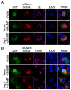
PRRSV Nsp5 blocks STING trafficking from the ER to the Golgi apparatus. (A and B) 3D4/21 cells were co-transfected with Nsp5 and STING expression plasmids for 24 h, and then treated with or without 2’3’-cGAMP for 12 h. Cells were fixed, permeabilized, and incubated with antibodies specific for Calreticulin (ER marker, A) and GORASP2 (Golgi marker, B), followed by counter-staining with DAPI. Samples were examined under a confocal microscope.
Figure 6.
Figure 6.
Porcine STING 1–153 amino acids is a critical region for interaction with Nsp5. (A) A schematic of porcine STING molecular structure and its truncated mutants. (B-E) HEK-293T cells were co-transfected with STING or its truncation mutants and Nsp5-GFP for 24 h, and the cells were harvested and subjected to Co-IP and subsequent Western blot analysis using the indicated antibodies. (F) mCherry-STING or its truncation mutants were co-transfected with Nsp5-GFP into 3D4/21 cells for 24 h. Cells were fixed, permeabilized, and examined for co-localization by confocal microscopy. The levels of co-localization between STING or its truncation mutants and Nsp5 were quantified by PCC using ImageJ software. Scale bars are10 µm.
Figure 7.
Figure 7.
The 36–47 and 58–67 amino acids of Nsp5 are the key regions for interaction with STING. (A) A schematic of PRRSV Nsp5 molecular structure and its truncation and deletion mutants. (B-F) Nsp5-GFP or its truncation mutants/deletion mutants were co-transfected with STING-HA in HEK-293T cells for 24 h, and the cells were harvested and subjected to Co-IP and subsequent Western blot analysis using indicated antibodies.
Figure 8.
Figure 8.
The regions of 36–47 and 58–67 amino acids of Nsp5 are crucial for interfering with STING signaling. (A) HEK-293T cells were co-transfected with TBK1-Myc together with mCherry-STING, Nsp5 or its deletion mutants for 24 h. (B) HEK-293T cells were co-transfected with IKKε-Myc together with mCherry-STING, Nsp5 or its deletion mutants for 24 h. Cell lysates were immunoprecipitated with anti-mCherry antibody and subjected to Co-IP and subsequent Western blot analysis. (C-D) 3D4/21 cells were transfected with Nsp5 or its deletion mutants for 24 h, and then stimulated with 2’3’-cGAMP for 12 h. Cell lysates were immunoprecipitated with anti-STING antibody and then analyzed by Western blotting for STING, TBK1, IKKε and GFP.
Figure 9.
Figure 9.
The regions of 36–47 and 58–67 amino acids of Nsp5 are crucial for blocking the STING trafficking and signaling. (A-B) 3D4/21 cells were co-transfected with STING and Nsp5 or Nsp5 deletion mutants and then treated with 2’3’-cGAMP. Cells were fixed, permeabilized and incubated with antibodies specific for calreticulin (A) and GORASP2 (B), followed by DAPI staining. Samples were examined under a confocal microscope. (C-D) HEK-293T cells were transfected with the ISRE-Luc (C), IFNβ-luc (D) reporter plasmid, together with Rluc reporter, cGAS, STING and Nsp5 or Nsp5 deletion mutant plasmids. Cells were collected 24 h after transfection, and ISRE/IFNβ promoter activity was evaluated via a luciferase reporter assay. * p < 0.05, ** p < 0.01.
Figure 10.
Figure 10.
Schematic model of the mechanism by which PRRSV Nsp5 inhibits the cGAS-STING signaling pathway, created in MedPeer. The interaction between PRRSV Nsp5 and STING hinders the transport of STING from ER to Golgi apparatus, and thus disrupts the assembly of the STING/TBK1/IKKε/IRF3 signaling complex and downstream antiviral IFN response.

References

    1. Lunney JK, Fang Y, Ladinig A, et al. Porcine reproductive and respiratory syndrome virus (PRRSV): pathogenesis and interaction with the immune system. Annu Rev Anim Biosci. 2016;4(1):129–16. doi: 10.1146/annurev-animal-022114-111025 - DOI - PubMed
    1. Guo Z, Chen XX, Li R, et al. The prevalent status and genetic diversity of porcine reproductive and respiratory syndrome virus in China: a molecular epidemiological perspective. Virol J. 2018;15(1):2. doi: 10.1186/s12985-017-0910-6 - DOI - PMC - PubMed
    1. Brinton MA, Gulyaeva AA, Balasuriya UBR, et al. ICTV virus taxonomy profile: arteriviridae 2021. J Gen Virol. 2021;102(8). doi: 10.1099/jgv.0.001632 - DOI - PMC - PubMed
    1. Kappes MA, Faaberg KS.. PRRSV structure, replication and recombination: origin of phenotype and genotype diversity. Virology. 2015;479–480:475–486. doi: 10.1016/j.virol.2015.02.012 - DOI - PMC - PubMed
    1. Zhang H, Sha H, Qin L, et al. Research progress in porcine reproductive and respiratory syndrome virus-host protein interactions. Animals (Basel). 2022;12(11):12. doi: 10.3390/ani12111381 - DOI - PMC - PubMed

MeSH terms

Substances

LinkOut - more resources