. 2025 Aug 20;8(3):e70106.
doi: 10.1002/jsp2.70106. eCollection 2025 Sep.

Wharton's Jelly Mesenchymal Stromal Cell-Derived Extracellular Vesicles Attenuate Intervertebral Disc Degeneration Under Inflammatory Stress in an In Vitro 3D Culture System

Affiliations

Wharton's Jelly Mesenchymal Stromal Cell-Derived Extracellular Vesicles Attenuate Intervertebral Disc Degeneration Under Inflammatory Stress in an In Vitro 3D Culture System

Veronica Tilotta et al. JOR Spine. .

Abstract

This study explores the therapeutic potential of extracellular vesicles (EVs) derived from Wharton's Jelly mesenchymal stem cells in an in vitro 3D model of intervertebral disc degeneration under inflammatory stress. The treatment with WJ-MSC-EVs enhanced nucleus pulposus cell proliferation, viability, and extracellular matrix synthesis while reducing oxidative stress and catabolic gene expression. These results support the promise of WJ-MSC-EVs as a novel, cell-free strategy for disc regeneration in inflammatory conditions.

Background: Extracellular vesicles (EVs) derived from mesenchymal stromal cells (MSCs) are emerging as a promising cell‐free strategy for intervertebral disc degeneration (IDD) treatment. This study aimed to evaluate the anabolic effect of Wharton's Jelly MSC (WJ‐MSC)‐derived EVs on degenerative human nucleus pulposus cells (hNPCs) under in vitro inflammation using a 3D culture model.

Methods: Following isolation, hNPCs (n = 10) were encapsulated in alginate beads and treated with 10, 50, and 100 μg/mL of WJ‐MSC‐EVs after preincubation with 10 ng/mL interleukin (IL)‐1β. Cell proliferation, viability, nitrite production, and glycosaminoglycan (GAG) content were assessed. Histological analyses evaluated extracellular matrix (ECM) production. Phenotypic (SOX9, KRT19), catabolic (MMP1, MMP13, ADAMTS5, IL6, NOS2), and anabolic (ACAN) ECM markers were analyzed by RT‐qPCR.

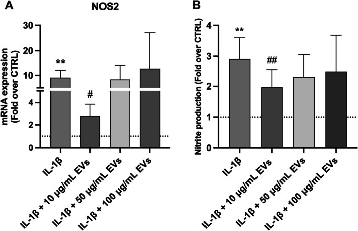
Results: WJ‐MSC‐EVs significantly promoted hNPC proliferation at all concentrations, with 10 μg/mL effectively counteracting IL‐1β catabolic effects. Live/dead staining showed reduced cell death in EV‐treated hNPCs compared to the IL‐1β‐only group. Nitrite production decreased after 7 days with 10 μg/mL WJ‐EVs, supported by reduced NOS2 expression. GAG content increased dose‐dependently, as confirmed by Alcian blue staining. WJ‐EVs positively modulated anabolic (ACAN, KRT19, SOX9), catabolic (ADAMTS5, MMP1, MMP13), and inflammatory (IL6) gene expression levels.

Conclusion: WJ‐MSC‐derived EVs demonstrate potential as a cell‐free therapeutic approach for IDD by enhancing hNPC growth, mitigating ECM degradation, and reducing oxidative stress‐related IDD progression. These findings warrant further investigation into the use of WJ‐EVs for IDD treatment.

Keywords: exosome; extracellular vesicles; intervertebral disc; intervertebral disc degeneration; intervertebral disc regeneration; low back pain; mesenchymal stromal cells.


Figures

FIGURE 1
FIGURE 1
Characterization of WJ‐MSC‐EVs. (A) Particle size distribution of WJ‐MSC‐EVs measured with NTA. (B) TEM showed the typical EV morphology in WJ‐MSC‐EVs (left, scale bar: 200 nm; right box, scale bar: 20 nm). (C) WB analysis of EV protein markers CD63, CD9, and TSG101. (D) Representative images of hNPCs incubated with PBS or PKH26‐labeled WJ‐MSC‐EVs (red). hNPC nuclei were stained with DAPI (blue). Magnification: 20× scale bar: 100 μm. Abbreviations: DAPI, 4′,6‐diamidino‐2‐phenylindole, EVs, extracellular vesicles, hNPCs, human nucleus pulposus cells, NTA, nanoparticle trafficking analysis, PBS, phosphate‐buffered saline, TEM, transmission electron microscopy, WB, Western blot, WJ‐MSCs, Wharton's Jelly mesenchymal stromal cells. Reproduced with permission from Tilotta et al. [14].
FIGURE 2
FIGURE 2
Characterization of WJ‐MSCs. (A) Surface markers (CD90, CD105, CD73, and SSEA3) detected through flow cytometric analysis. (B) Multilineage differentiation of WJ‐MSCs towards the osteogenic, adipogenic, and chondrogenic lineages was confirmed by Alizarin Red, Oil red O, and Alcian Blue staining (left and center, scale bars: 50 μm; right, scale bar: 100 μm). Abbreviation: WJ‐MSCs, Wharton's Jelly mesenchymal stromal cells. Reproduced with permission from Tilotta et al. [14].
FIGURE 3
FIGURE 3
WJ‐MSC‐derived EVs promoted hNPC proliferation and viability. (A) Cell content significantly increased after treatment with 10 μg/mL WJ‐MSC‐EVs at day 4, 10, and 14, as compared with the IL‐1β control group (n = 4). Data were analyzed through two‐way ANOVA with Dunnett's multiple comparisons test, where each group was compared with IL‐1β. Within each timepoint, values were analyzed using unpaired t tests with a false discovery rate approach. (B) After 24 h, the WJ‐MSC‐EV treatment significantly reduced IL‐1β‐pre‐treated hNPC death at 100 μg/mL at flow cytometry (n = 4). Data were analyzed through two‐way ANOVA with Dunnett's multiple comparisons test, where each group was compared with IL‐1β. (C) Live/Dead staining showed reduced cell death in hNPCs treated with 10 and 100 μg/mL WJ‐MSC‐EVs (n = 4). Magnification: 20×, scale bar: 100 μm.*p < 0.05, **p < 0.01, compared to the control group. # p < 0.05, ## p < 0.01, ### p < 0.001 compared to the IL‐1β group. Abbreviations: AM, acetoxymethylester; hNPCs, human nucleus pulposus cells; WJ‐MSC‐EVs, Wharton's Jelly mesenchymal stromal cell‐derived extracellular vesicles.
FIGURE 4
FIGURE 4
(A) NOS2 mRNA levels were significantly reduced by 10 μg/mL WJ‐MSC‐EVs (n = 5). EVs led to a significant decrease in nitrite release in cell supernatant (B) after 7 days in groups treated with 10 μg/mL EVs compared to the IL‐1β group (n = 6). Data were analyzed through one‐way ANOVA with Dunnett's multiple comparisons test, where each group was compared with IL‐1β. **p < 0.01 compared to the control group, # p < 0.05, ## p < 0.01 compared to the IL‐1β group. Abbreviations: HNPCs, human nucleus pulposus cells; NOS2, nitric oxide synthase 2; WJ‐MSC‐EVs, Wharton's Jelly mesenchymal stromal cell‐derived extracellular vesicles.
FIGURE 5
FIGURE 5
WJ‐MSC‐EVs enhanced ECM synthesis in IL‐1β‐treated hNPCs. (A) GAG/DNA ratio in hNPCs after WJ‐MSC‐EVs treatment demonstrated an increase in all experimental groups, although significant only in the 10 μg/mL WJ‐MSC‐EV group. Data were analyzed through one‐way ANOVA with Dunnett's multiple comparisons test, where each group was compared with IL‐1β. (B) Representative images of Alcian Blue staining of hNPC alginate beads are shown. Blue color indicates proteoglycans. Arrowheads point to proteoglycan deposits. Upper rows, scale bar: 500 μm; bottom boxes, scale bar: 100 μm. Data are expressed as GAG/DNA ratio percent variation between the control and experimental groups (n = 5). *p < 0.05, ***p < 0.001 compared to the control group, ##p < 0.01 compared to the IL‐1β group. Abbreviations: GAG, glycosaminoglycans; hNPCs, human nucleus pulposus cells; WJ‐MSC‐EVs, Wharton's Jelly mesenchymal stromal cell‐derived extracellular vesicles.
FIGURE 6
FIGURE 6
WJ‐MSC‐EVs maintained the hNPC phenotype and reduced catabolic gene expression levels. WJ‐MSC‐EV treatment resulted in a significant decrease of ADAMTS5 (A), MMP1 (B), MMP13 (C) and IL6 (D) mRNA levels, while increasing ACAN (E), SOX9 (F) and KRT19 (G) gene expression. Results were normalized based on GAPDH expression and calculated as fold change compared to the controls. Data were analyzed through one‐way ANOVA with Dunnett's multiple comparisons test, where each group was compared with IL‐1β. *p < 0.05, ***p < 0.001, ****p < 0.0001 compared to the control group, # p < 0.05, ## p < 0.01 compared to the IL1‐β group. Abbreviations: ACAN, aggrecan; ADAMTS, a disintegrin and metalloproteinase with thrombospondin motifs; hNPCs, human nucleus pulposus cells; IL, interleukin; MMP, matrix metalloproteinase; NOS, nitric oxide synthase; SOX, SRY‐box transcription factor; WJ‐MSC‐EVs = Wharton's Jelly mesenchymal stromal cell‐derived extracellular vesicles.

References

    1. Ferreira M. L., de Luca K., Haile L. M., et al., “Global, Regional, and National Burden of Low Back Pain, 1990–2020, Its Attributable Risk Factors, and Projections to 2050: A Systematic Analysis of the Global Burden of Disease Study 2021,” Lancet Rheumatology 5 (2023): e316–e329. - PMC - PubMed
    1. Ambrosio L., Mazzuca G., Maguolo A., et al., “The Burden of Low Back Pain in Children and Adolescents With Overweight and Obesity: From Pathophysiology to Prevention and Treatment Strategies,” Therapeutic Advances in Musculoskeletal Disease 15 (2023): 1759720X231188831. - PMC - PubMed
    1. Lyu F.‐J., Cui H., Pan H., et al., “Painful Intervertebral Disc Degeneration and Inflammation: From Laboratory Evidence to Clinical Interventions,” Bone Research 9 (2021): 7. - PMC - PubMed
    1. Ambrosio L., Schol J., Ruiz‐Fernández C., et al., “Getting to the Core: Exploring the Embryonic Development From Notochord to Nucleus Pulposus,” Journal of Developmental Biology 12 (2024): 18. - PMC - PubMed
    1. Vo N. V., Hartman R. A., Patil P. R., et al., “Molecular Mechanisms of Biological Aging in Intervertebral Discs,” Journal of Orthopaedic Research 34 (2016): 1289–1306. - PMC - PubMed

LinkOut - more resources